 Movie
Movie Controller

[English] 日本語
 Yorodumi
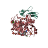
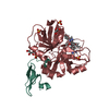
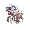
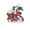
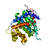
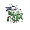
- PDB-5l30: Factor VIIa in complex with the inhibitor (2R,15R)-2-[(1-aminoiso... -
+ Open data

- Basic information
| Entry | Database: PDB / ID: 5l30 | ||||||
|---|---|---|---|---|---|---|---|
| Title | Factor VIIa in complex with the inhibitor (2R,15R)-2-[(1-aminoisoquinolin-6-yl)amino]-4,15,17-trimethyl-7-[1-(1H-tetrazol-5-yl)cyclopropyl]-13-oxa-4,11-diazatricyclo[14.2.2.1~6,10~]henicosa-1(18),6(21),7,9,16,19-hexaene-3,12-dione | ||||||
 Components Components | (Coagulation factor VII ...) x 2 | ||||||
 Keywords Keywords | Hydrolase/Hydrolase Inhibitor / Glycoprotein / hydrolase / serine protease / plasma / blood coagulation factor / protein inhibitor complex / calcium-binding / Hydrolase-Hydrolase Inhibitor complex | ||||||
| Function / homology |  Function and homology information Function and homology informationcoagulation factor VIIa / response to Thyroid stimulating hormone / response to astaxanthin / response to thyrotropin-releasing hormone / response to 2,3,7,8-tetrachlorodibenzodioxine / response to carbon dioxide / serine-type peptidase complex / response to genistein / response to vitamin K / positive regulation of platelet-derived growth factor receptor signaling pathway ...coagulation factor VIIa / response to Thyroid stimulating hormone / response to astaxanthin / response to thyrotropin-releasing hormone / response to 2,3,7,8-tetrachlorodibenzodioxine / response to carbon dioxide / serine-type peptidase complex / response to genistein / response to vitamin K / positive regulation of platelet-derived growth factor receptor signaling pathway / positive regulation of leukocyte chemotaxis / response to thyroxine / response to cholesterol / positive regulation of positive chemotaxis / : / response to growth hormone / positive regulation of blood coagulation / animal organ regeneration / positive regulation of TOR signaling / Transport of gamma-carboxylated protein precursors from the endoplasmic reticulum to the Golgi apparatus / Gamma-carboxylation of protein precursors / Removal of aminoterminal propeptides from gamma-carboxylated proteins / BMAL1:CLOCK,NPAS2 activates circadian expression / serine-type peptidase activity / circadian rhythm / protein processing / response to estrogen / Golgi lumen / blood coagulation / response to estradiol / extracellular matrix / vesicle / response to hypoxia / positive regulation of cell migration / endoplasmic reticulum lumen / signaling receptor binding / serine-type endopeptidase activity / calcium ion binding / : / extracellular region / plasma membrane Similarity search - Function | ||||||
| Biological species |  Homo sapiens (human) Homo sapiens (human) | ||||||
| Method |  X-RAY DIFFRACTION / SYNCHROTRON / MOLECULAR REPLACEMENT / Resolution: 1.73 Å X-RAY DIFFRACTION / SYNCHROTRON / MOLECULAR REPLACEMENT / Resolution: 1.73 Å | ||||||
 Authors Authors | Wei, A. | ||||||
 Citation Citation | Journal: Bioorg.Med.Chem.Lett. / Year: 2016 Title: Synthesis and P1' SAR exploration of potent macrocyclic tissue factor-factor VIIa inhibitors. Authors: Ladziata, V.U. / Glunz, P.W. / Zou, Y. / Zhang, X. / Jiang, W. / Jacutin-Porte, S. / Cheney, D.L. / Wei, A. / Luettgen, J.M. / Harper, T.M. / Wong, P.C. / Seiffert, D. / Wexler, R.R. / Priestley, E.S. | ||||||
| History |
|
- Structure visualization
| Structure viewer | Molecule: MolmilJmol/JSmol |
|---|
- Downloads & links
-Download
| PDBx/mmCIF format | 5l30.cif.gz | 90.6 KB | Display | PDBx/mmCIF format |
|---|---|---|---|---|
| PDB format | pdb5l30.ent.gz | 65.4 KB | Display | PDB format |
| PDBx/mmJSON format | 5l30.json.gz | Tree view | PDBx/mmJSON format | |
| Others |  Other downloads Other downloads |
-Validation report
| Arichive directory | https://data.pdbj.org/pub/pdb/validation_reports/l3/5l30ftp://data.pdbj.org/pub/pdb/validation_reports/l3/5l30 | HTTPS FTP |
|---|
-Related structure data
| Related structure data | 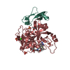 5l2yC  5l2zC  1danS S: Starting model for refinement C: citing same article ( |
|---|---|
| Similar structure data |


-Links
 PDBj
PDBj


- Assembly
| Deposited unit | 
| ||||||||
|---|---|---|---|---|---|---|---|---|---|
| 1 |
| ||||||||
| Unit cell |
| ||||||||
| Components on special symmetry positions |
|
-Components
-Coagulation factor VII ... , 2 types, 2 molecules HL
| #1: Protein | Mass: 28103.256 Da / Num. of mol.: 1 Source method: isolated from a genetically manipulated source Source: (gene. exp.) Homo sapiens (human) / Gene: F7 / Production host:  Cricetinae (hamsters) / References: UniProt: P08709, coagulation factor VIIa Cricetinae (hamsters) / References: UniProt: P08709, coagulation factor VIIa |
|---|---|
| #2: Protein | Mass: 6387.201 Da / Num. of mol.: 1 Source method: isolated from a genetically manipulated source Source: (gene. exp.) Homo sapiens (human) / Gene: F7 / Production host: Cricetinae (hamsters) / References: UniProt: P08709, coagulation factor VIIa |
-Non-polymers , 5 types, 418 molecules 


| #3: Chemical | ChemComp-70A / ( Mass: 617.700 Da / Num. of mol.: 1 / Source method: obtained synthetically / Formula: C34H35N9O3 Mass: 617.700 Da / Num. of mol.: 1 / Source method: obtained synthetically / Formula: C34H35N9O3 | ||||
|---|---|---|---|---|---|
| #4: Chemical | ChemComp-CA /  Mass: 40.078 Da / Num. of mol.: 1 / Source method: obtained synthetically / Formula: Ca Mass: 40.078 Da / Num. of mol.: 1 / Source method: obtained synthetically / Formula: Ca | ||||
| #5: Chemical | ChemComp-SO4 /  Mass: 96.063 Da / Num. of mol.: 4 / Source method: obtained synthetically / Formula: SO4 Mass: 96.063 Da / Num. of mol.: 4 / Source method: obtained synthetically / Formula: SO4#6: Chemical |  Mass: 92.094 Da / Num. of mol.: 2 / Source method: obtained synthetically / Formula: C3H8O3 Mass: 92.094 Da / Num. of mol.: 2 / Source method: obtained synthetically / Formula: C3H8O3#7: Water | ChemComp-HOH / | Mass: 18.015 Da / Num. of mol.: 410 / Source method: isolated from a natural source / Formula: H2O |
-Details
| Has protein modification | Y |
|---|
-Experimental details
-Experiment
| Experiment | Method: X-RAY DIFFRACTION / Number of used crystals: 1 |
|---|
- Sample preparation
| Crystal | Density Matthews: 3.77 Å3/Da / Density % sol: 67.38 % |
|---|---|
| Crystal grow | Temperature: 277 K / Method: vapor diffusion, hanging drop / pH: 6 Details: 100 mM MES, pH 6.0, 20 mM CACL2, 17.5%(W/V) PEG 6000 |
-Data collection
| Diffraction | Mean temperature: 100 K |
|---|---|
| Diffraction source | Source: SYNCHROTRON / Site: APS  / Beamline: 17-BM / Wavelength: 1 Å / Beamline: 17-BM / Wavelength: 1 Å |
| Detector | Type: ADSC QUANTUM 210 / Detector: CCD / Date: Nov 10, 2008 |
| Radiation | Protocol: SINGLE WAVELENGTH / Monochromatic (M) / Laue (L): M / Scattering type: x-ray |
| Radiation wavelength | Wavelength: 1 Å / Relative weight: 1 |
| Reflection | Resolution: 1.73→50 Å / Num. obs: 55544 / % possible obs: 99.8 % / Observed criterion σ(I): 0 / Redundancy: 10.5 % / Biso Wilson estimate: 25.66 Å2 / Rsym value: 0.056 / Net I/σ(I): 42.8 |
| Reflection shell | Resolution: 1.73→1.79 Å / Redundancy: 10.2 % / Rmerge(I) obs: 0.625 / Mean I/σ(I) obs: 4.8 / Rejects: 0 / % possible all: 99.7 |
- Processing
| Software |
| ||||||||||||||||||||||||||||||||||||||||||||||||||||||||||||||||||||||||||||||||||||||||||||||||||||||||||||
|---|---|---|---|---|---|---|---|---|---|---|---|---|---|---|---|---|---|---|---|---|---|---|---|---|---|---|---|---|---|---|---|---|---|---|---|---|---|---|---|---|---|---|---|---|---|---|---|---|---|---|---|---|---|---|---|---|---|---|---|---|---|---|---|---|---|---|---|---|---|---|---|---|---|---|---|---|---|---|---|---|---|---|---|---|---|---|---|---|---|---|---|---|---|---|---|---|---|---|---|---|---|---|---|---|---|---|---|---|---|
| Refinement | Method to determine structure: MOLECULAR REPLACEMENT Starting model: 1DAN Resolution: 1.73→25.33 Å / Cor.coef. Fo:Fc: 0.955 / Cor.coef. Fo:Fc free: 0.948 / Rfactor Rfree error: 0 / SU R Cruickshank DPI: 0.08 / Cross valid method: THROUGHOUT / σ(F): 0 / SU R Blow DPI: 0.089 / SU Rfree Blow DPI: 0.082 / SU Rfree Cruickshank DPI: 0.077
| ||||||||||||||||||||||||||||||||||||||||||||||||||||||||||||||||||||||||||||||||||||||||||||||||||||||||||||
| Displacement parameters | Biso max: 114.26 Å2 / Biso mean: 29.4 Å2 / Biso min: 13.5 Å2
| ||||||||||||||||||||||||||||||||||||||||||||||||||||||||||||||||||||||||||||||||||||||||||||||||||||||||||||
| Refine analyze | Luzzati coordinate error obs: 0.22 Å | ||||||||||||||||||||||||||||||||||||||||||||||||||||||||||||||||||||||||||||||||||||||||||||||||||||||||||||
| Refinement step | Cycle: final / Resolution: 1.73→25.33 Å
| ||||||||||||||||||||||||||||||||||||||||||||||||||||||||||||||||||||||||||||||||||||||||||||||||||||||||||||
| Refine LS restraints |
| ||||||||||||||||||||||||||||||||||||||||||||||||||||||||||||||||||||||||||||||||||||||||||||||||||||||||||||
| LS refinement shell | Resolution: 1.73→1.77 Å / Total num. of bins used: 20
|
