 Movie
Movie Controller

[English] 日本語
 Yorodumi
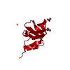
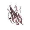
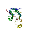
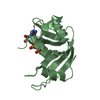
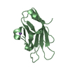
- PDB-2i89: Structure of septuple mutant of Rat Outer Mitochondrial Membrane ... -
+ Open data

- Basic information
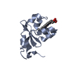
| Entry | Database: PDB / ID: 2i89 | ||||||
|---|---|---|---|---|---|---|---|
| Title | Structure of septuple mutant of Rat Outer Mitochondrial Membrane Cytochrome B5 | ||||||
 Components Components | Cytochrome b5 type B | ||||||
 Keywords Keywords | ELECTRON TRANSPORT / CYTOCHROME B5 / HEME | ||||||
| Function / homology |  Function and homology information Function and homology informationSphingolipid de novo biosynthesis / eNOS activation / Phase I - Functionalization of compounds / nitric-oxide synthase complex / quinol-cytochrome-c reductase activity / nitric oxide biosynthetic process / enzyme activator activity / mitochondrial outer membrane / heme binding / metal ion binding Similarity search - Function | ||||||
| Biological species |  | ||||||
| Method |  X-RAY DIFFRACTION / FOURIER SYNTHESIS / Resolution: 2.1 Å X-RAY DIFFRACTION / FOURIER SYNTHESIS / Resolution: 2.1 Å | ||||||
 Authors Authors | Terzyan, S. / Zhang, X.C. / Benson, D.R. / Wang, L. / Sun, N. | ||||||
 Citation Citation | Journal: Biochemistry / Year: 2006 Title: A histidine/tryptophan pi-stacking interaction stabilizes the heme-independent folding core of microsomal apocytochrome b5 relative to that of mitochondrial apocytochrome b5. Authors: Wang, L. / Sun, N. / Terzyan, S. / Zhang, X. / Benson, D.R. | ||||||
| History |
|
- Structure visualization
| Structure viewer | Molecule: MolmilJmol/JSmol |
|---|
- Downloads & links
-Download
| PDBx/mmCIF format | 2i89.cif.gz | 97.9 KB | Display | PDBx/mmCIF format |
|---|---|---|---|---|
| PDB format | pdb2i89.ent.gz | 76.3 KB | Display | PDB format |
| PDBx/mmJSON format | 2i89.json.gz | Tree view | PDBx/mmJSON format | |
| Others |  Other downloads Other downloads |
-Validation report
| Arichive directory | https://data.pdbj.org/pub/pdb/validation_reports/i8/2i89ftp://data.pdbj.org/pub/pdb/validation_reports/i8/2i89 | HTTPS FTP |
|---|
-Related structure data
| Related structure data | 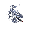 1iccS S: Starting model for refinement |
|---|---|
| Similar structure data |


-Links
 PDBj
PDBj
- Assembly
| Deposited unit | 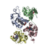
| ||||||||
|---|---|---|---|---|---|---|---|---|---|
| 1 | 
| ||||||||
| 2 | 
| ||||||||
| 3 | 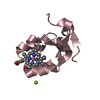
| ||||||||
| 4 | 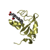
| ||||||||
| 5 | 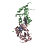
| ||||||||
| Unit cell |
|
-Components
| #1: Protein | Mass: 10494.436 Da / Num. of mol.: 4 / Fragment: Water Soluble Domain / Mutation: R15H/A18S/E20S/I25L/I32L/L47R/L71S Source method: isolated from a genetically manipulated source Source: (gene. exp.)  #2: Chemical |   Mass: 24.305 Da / Num. of mol.: 3 / Source method: obtained synthetically / Formula: Mg Mass: 24.305 Da / Num. of mol.: 3 / Source method: obtained synthetically / Formula: Mg#3: Chemical | ChemComp-HEM /   Mass: 616.487 Da / Num. of mol.: 4 / Source method: obtained synthetically / Formula: C34H32FeN4O4 Mass: 616.487 Da / Num. of mol.: 4 / Source method: obtained synthetically / Formula: C34H32FeN4O4#4: Water | ChemComp-HOH / |  Mass: 18.015 Da / Num. of mol.: 212 / Source method: isolated from a natural source / Formula: H2O Mass: 18.015 Da / Num. of mol.: 212 / Source method: isolated from a natural source / Formula: H2O |
|---|
-Experimental details
-Experiment
| Experiment | Method: X-RAY DIFFRACTION / Number of used crystals: 1 |
|---|
- Sample preparation
| Crystal | Density Matthews: 2.11 Å3/Da / Density % sol: 41.66 % |
|---|---|
| Crystal grow | Temperature: 278 K / pH: 6.8 Details: PEG8K 30%, O.2M MgAc, 0.1M Pipes, VAPOR DIFFUSION, HANGING DROP, temperature 278K, pH 6.80 |
-Data collection
| Diffraction | Mean temperature: 100 K |
|---|---|
| Diffraction source | Source: ROTATING ANODE / Type: RIGAKU / Wavelength: 1.5418 |
| Detector | Type: MAR scanner 345 mm plate / Detector: IMAGE PLATE / Date: May 20, 2004 / Details: OSMIC BLUE OPTICS |
| Radiation | Monochromator: OSMIC MIRRORS / Protocol: SINGLE WAVELENGTH / Monochromatic (M) / Laue (L): M / Scattering type: x-ray |
| Radiation wavelength | Wavelength: 1.5418 Å / Relative weight: 1 |
| Reflection | Resolution: 2.1→50 Å / Num. obs: 19485 / % possible obs: 92.6 % / Observed criterion σ(I): -3 / Redundancy: 3.6 % / Biso Wilson estimate: 29.98 Å2 / Rmerge(I) obs: 0.099 / Net I/σ(I): 9.3 |
| Reflection shell | Resolution: 2.1→2.18 Å / Rmerge(I) obs: 0.476 / Mean I/σ(I) obs: 2.6 / % possible all: 90.4 |
- Processing
| Software |
| ||||||||||||||||||||||||||||||||||||||||||||||||||||||||||||||||||||||||||||||||
|---|---|---|---|---|---|---|---|---|---|---|---|---|---|---|---|---|---|---|---|---|---|---|---|---|---|---|---|---|---|---|---|---|---|---|---|---|---|---|---|---|---|---|---|---|---|---|---|---|---|---|---|---|---|---|---|---|---|---|---|---|---|---|---|---|---|---|---|---|---|---|---|---|---|---|---|---|---|---|---|---|---|
| Refinement | Method to determine structure: FOURIER SYNTHESIS Starting model: 1ICC Resolution: 2.1→50 Å / Isotropic thermal model: ISOTROPIC / Cross valid method: THROUGHOUT / σ(F): 0 / Stereochemistry target values: ENGH & HUBER
| ||||||||||||||||||||||||||||||||||||||||||||||||||||||||||||||||||||||||||||||||
| Solvent computation | Bsol: 50.02 Å2 / ksol: 0.375 e/Å3 | ||||||||||||||||||||||||||||||||||||||||||||||||||||||||||||||||||||||||||||||||
| Displacement parameters | Biso mean: 29.67 Å2
| ||||||||||||||||||||||||||||||||||||||||||||||||||||||||||||||||||||||||||||||||
| Refinement step | Cycle: LAST / Resolution: 2.1→50 Å
| ||||||||||||||||||||||||||||||||||||||||||||||||||||||||||||||||||||||||||||||||
| Refine LS restraints |
| ||||||||||||||||||||||||||||||||||||||||||||||||||||||||||||||||||||||||||||||||
| Xplor file |
|
