 ムービー
ムービー コントローラー


+ データを開く

- 基本情報
基本情報
| 登録情報 | データベース: PDB / ID: 4l1d | ||||||
|---|---|---|---|---|---|---|---|
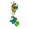
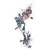
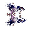
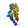
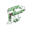
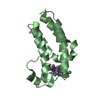
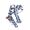
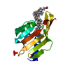
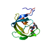
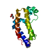
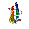
| タイトル | Voltage-gated sodium channel beta3 subunit Ig domain | ||||||
 要素 要素 | Sodium channel subunit beta-3 | ||||||
 キーワード キーワード | MEMBRANE PROTEIN / V-type immunoglobulin fold / sodium channel / voltage-gated / sodium channel alpha subunit / membrane | ||||||
| 機能・相同性 |  機能・相同性情報 機能・相同性情報regulation of ventricular cardiac muscle cell membrane depolarization / SA node cell action potential / voltage-gated sodium channel activity involved in cardiac muscle cell action potential / atrial cardiac muscle cell action potential / regulation of atrial cardiac muscle cell membrane depolarization / membrane depolarization during cardiac muscle cell action potential / positive regulation of sodium ion transport / membrane depolarization during action potential / ventricular cardiac muscle cell action potential / cardiac muscle cell action potential involved in contraction ...regulation of ventricular cardiac muscle cell membrane depolarization / SA node cell action potential / voltage-gated sodium channel activity involved in cardiac muscle cell action potential / atrial cardiac muscle cell action potential / regulation of atrial cardiac muscle cell membrane depolarization / membrane depolarization during cardiac muscle cell action potential / positive regulation of sodium ion transport / membrane depolarization during action potential / ventricular cardiac muscle cell action potential / cardiac muscle cell action potential involved in contraction / sodium channel inhibitor activity / voltage-gated sodium channel complex / Interaction between L1 and Ankyrins / sodium ion transport / Phase 0 - rapid depolarisation / regulation of heart rate by cardiac conduction / sodium channel regulator activity / membrane depolarization / positive regulation of heart rate / cardiac muscle contraction / protein localization to plasma membrane / sodium ion transmembrane transport / Z disc / nervous system development / transmembrane transporter binding / membrane / plasma membrane / cytosol 類似検索 - 分子機能 | ||||||
| 生物種 |  Homo sapiens (ヒト) Homo sapiens (ヒト) | ||||||
| 手法 |  X線回折 / シンクロトロン / 分子置換 / 解像度: 2.5 Å X線回折 / シンクロトロン / 分子置換 / 解像度: 2.5 Å | ||||||
 データ登録者 データ登録者 | Namadurai, S. / Weimhofer, M. / Rajappa, R. / Stott, K. / Klingauf, J. / Chirgadze, D.Y. / Jackson, A.P. | ||||||
 引用 引用 | ジャーナル: J.Biol.Chem. / 年: 2014 タイトル: Crystal Structure and Molecular Imaging of the Nav Channel beta 3 Subunit Indicates a Trimeric Assembly. 著者: Namadurai, S. / Balasuriya, D. / Rajappa, R. / Wiemhofer, M. / Stott, K. / Klingauf, J. / Edwardson, J.M. / Chirgadze, D.Y. / Jackson, A.P. | ||||||
| 履歴 |
|
- 構造の表示
構造の表示
| 構造ビューア | 分子: MolmilJmol/JSmol |
|---|
- ダウンロードとリンク
-ダウンロード
| PDBx/mmCIF形式 | 4l1d.cif.gz | 147 KB | 表示 | PDBx/mmCIF形式 |
|---|---|---|---|---|
| PDB形式 | pdb4l1d.ent.gz | 118 KB | 表示 | PDB形式 |
| PDBx/mmJSON形式 | 4l1d.json.gz | ツリー表示 | PDBx/mmJSON形式 | |
| その他 |  その他のダウンロード その他のダウンロード |
-検証レポート
| アーカイブディレクトリ | https://data.pdbj.org/pub/pdb/validation_reports/l1/4l1dftp://data.pdbj.org/pub/pdb/validation_reports/l1/4l1d | HTTPS FTP |
|---|
-関連構造データ


-リンク
 PDBj
PDBj


- 集合体
集合体
| 登録構造単位 | 
| ||||||||||||||||||||||||||||||||||||
|---|---|---|---|---|---|---|---|---|---|---|---|---|---|---|---|---|---|---|---|---|---|---|---|---|---|---|---|---|---|---|---|---|---|---|---|---|---|
| 1 | 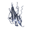
| ||||||||||||||||||||||||||||||||||||
| 2 | 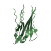
| ||||||||||||||||||||||||||||||||||||
| 3 | 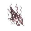
| ||||||||||||||||||||||||||||||||||||
| 単位格子 |
| ||||||||||||||||||||||||||||||||||||
| Components on special symmetry positions |
| ||||||||||||||||||||||||||||||||||||
| 非結晶学的対称性 (NCS) | NCSドメイン:
NCSドメイン領域:
|
-要素
| #1: タンパク質 | 分子量: 14832.702 Da / 分子数: 3 / 断片: Ig domain, UNP residues 25-145 / 由来タイプ: 組換発現 / 由来: (組換発現) Homo sapiens (ヒト) / 遺伝子: KIAA1158, SCN3B / プラスミド: pTT3 / 細胞株 (発現宿主): HEK293F / 発現宿主: Homo sapiens (ヒト) / 参照: UniProt: Q9NY72#2: 水 | ChemComp-HOH / |  分子量: 18.015 Da / 分子数: 117 / 由来タイプ: 天然 / 式: H2O 分子量: 18.015 Da / 分子数: 117 / 由来タイプ: 天然 / 式: H2OHas protein modification | Y | |
|---|
-実験情報
-実験
| 実験 | 手法: X線回折 / 使用した結晶の数: 1 |
|---|
- 試料調製
試料調製
| 結晶 | マシュー密度: 2.46 Å3/Da / 溶媒含有率: 50 % |
|---|---|
| 結晶化 | 温度: 292 K / 手法: 蒸気拡散法, シッティングドロップ法 / pH: 9.5 詳細: 1M Sodium citrate tribasic, 0.1M CHES/Sodium hydroxide pH9.5, VAPOR DIFFUSION, SITTING DROP, temperature 292K |
-データ収集
| 回折 | 平均測定温度: 100 K |
|---|---|
| 放射光源 | 由来: シンクロトロン / サイト: Diamond  / ビームライン: I04 / 波長: 0.9795 Å / ビームライン: I04 / 波長: 0.9795 Å |
| 検出器 | タイプ: ADSC QUANTUM 315 / 検出器: CCD / 日付: 2012年8月14日 |
| 放射 | モノクロメーター: double crystal Si(111) / プロトコル: SINGLE WAVELENGTH / 単色(M)・ラウエ(L): M / 散乱光タイプ: x-ray |
| 放射波長 | 波長: 0.9795 Å / 相対比: 1 |
| 反射 | 解像度: 2.5→30 Å / Num. all: 15706 / Num. obs: 15675 / % possible obs: 99.8 % / Observed criterion σ(F): 0 / Observed criterion σ(I): 0 / 冗長度: 5.5 % / Biso Wilson estimate: 52.6 Å2 / Rmerge(I) obs: 0.076 / Rsym value: 0.076 / Net I/σ(I): 13.8 |
| 反射 シェル | 解像度: 2.5→2.64 Å / 冗長度: 5.7 % / Rmerge(I) obs: 0.633 / Mean I/σ(I) obs: 2.8 / Num. unique all: 2257 / Rsym value: 0.633 / % possible all: 100 |
- 解析
解析
| ソフトウェア |
| ||||||||||||||||||||||||||||||||||||||||||||||||||||||||||||||||||||||||||||||||||||||||||||||||||||
|---|---|---|---|---|---|---|---|---|---|---|---|---|---|---|---|---|---|---|---|---|---|---|---|---|---|---|---|---|---|---|---|---|---|---|---|---|---|---|---|---|---|---|---|---|---|---|---|---|---|---|---|---|---|---|---|---|---|---|---|---|---|---|---|---|---|---|---|---|---|---|---|---|---|---|---|---|---|---|---|---|---|---|---|---|---|---|---|---|---|---|---|---|---|---|---|---|---|---|---|---|---|
| 精密化 | 構造決定の手法: 分子置換 開始モデル: PDB ENTRY 2X1X, 1I8L AND 1F97 解像度: 2.5→30 Å / SU ML: 0.28 / 交差検証法: THROUGHOUT / σ(F): 1.45 / 位相誤差: 23.97 / 立体化学のターゲット値: ML
| ||||||||||||||||||||||||||||||||||||||||||||||||||||||||||||||||||||||||||||||||||||||||||||||||||||
| 溶媒の処理 | 減衰半径: 0.9 Å / VDWプローブ半径: 1.11 Å / 溶媒モデル: FLAT BULK SOLVENT MODEL | ||||||||||||||||||||||||||||||||||||||||||||||||||||||||||||||||||||||||||||||||||||||||||||||||||||
| 精密化ステップ | サイクル: LAST / 解像度: 2.5→30 Å
| ||||||||||||||||||||||||||||||||||||||||||||||||||||||||||||||||||||||||||||||||||||||||||||||||||||
| 拘束条件 |
| ||||||||||||||||||||||||||||||||||||||||||||||||||||||||||||||||||||||||||||||||||||||||||||||||||||
| Refine LS restraints NCS |
| ||||||||||||||||||||||||||||||||||||||||||||||||||||||||||||||||||||||||||||||||||||||||||||||||||||
| LS精密化 シェル |
| ||||||||||||||||||||||||||||||||||||||||||||||||||||||||||||||||||||||||||||||||||||||||||||||||||||
| 精密化 TLS | 手法: refined / Refine-ID: X-RAY DIFFRACTION
| ||||||||||||||||||||||||||||||||||||||||||||||||||||||||||||||||||||||||||||||||||||||||||||||||||||
| 精密化 TLSグループ |
|
