 Movie
Movie Controller


+ Open data

- Basic information
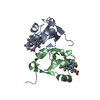
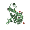
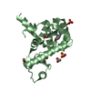
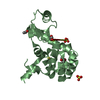
| Entry | Database: PDB / ID: 1awp | ||||||
|---|---|---|---|---|---|---|---|
| Title | RAT OUTER MITOCHONDRIAL MEMBRANE CYTOCHROME B5 | ||||||
 Components Components | CYTOCHROME B5 | ||||||
 Keywords Keywords | ELECTRON TRANSPORT / CYTOCHROME / HEME | ||||||
| Function / homology |  Function and homology information Function and homology informationSphingolipid de novo biosynthesis / eNOS activation / Phase I - Functionalization of compounds / nitric-oxide synthase complex / quinol-cytochrome-c reductase activity / nitric oxide biosynthetic process / enzyme activator activity / mitochondrial outer membrane / heme binding / metal ion binding Similarity search - Function | ||||||
| Biological species |  | ||||||
| Method |  X-RAY DIFFRACTION / MOLECULAR REPLACEMENT / Resolution: 2 Å X-RAY DIFFRACTION / MOLECULAR REPLACEMENT / Resolution: 2 Å | ||||||
 Authors Authors | Wang, X. / Zhang, X. | ||||||
 Citation Citation | Journal: Biochemistry / Year: 1998 Title: The reduction potential of cytochrome b5 is modulated by its exposed heme edge. Authors: Rivera, M. / Seetharaman, R. / Girdhar, D. / Wirtz, M. / Zhang, X. / Wang, X. / White, S. | ||||||
| History |
|
- Structure visualization
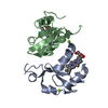
| Structure viewer | Molecule: MolmilJmol/JSmol |
|---|
- Downloads & links
-Download
| PDBx/mmCIF format | 1awp.cif.gz | 51.6 KB | Display | PDBx/mmCIF format |
|---|---|---|---|---|
| PDB format | pdb1awp.ent.gz | 37.3 KB | Display | PDB format |
| PDBx/mmJSON format | 1awp.json.gz | Tree view | PDBx/mmJSON format | |
| Others |  Other downloads Other downloads |
-Validation report
| Arichive directory | https://data.pdbj.org/pub/pdb/validation_reports/aw/1awpftp://data.pdbj.org/pub/pdb/validation_reports/aw/1awp | HTTPS FTP |
|---|
-Related structure data
| Related structure data | 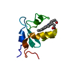 1b5mS S: Starting model for refinement |
|---|---|
| Similar structure data |

-Links
 PDBj
PDBj
- Assembly
| Deposited unit | 
| ||||||||
|---|---|---|---|---|---|---|---|---|---|
| 1 |
| ||||||||
| Unit cell |
| ||||||||
| Noncrystallographic symmetry (NCS) | NCS oper: (Code: given Matrix: (-0.54813, 0.5997, 0.58302), Vector: |
-Components
| #1: Protein | Mass: 10419.405 Da / Num. of mol.: 2 / Fragment: WATER SOLUBLE DOMAIN / Mutation: V45L, V61L Source method: isolated from a genetically manipulated source Source: (gene. exp.)  #2: Chemical |   Mass: 616.487 Da / Num. of mol.: 2 / Source method: obtained synthetically / Formula: C34H32FeN4O4 Mass: 616.487 Da / Num. of mol.: 2 / Source method: obtained synthetically / Formula: C34H32FeN4O4#3: Water | ChemComp-HOH / |  Mass: 18.015 Da / Num. of mol.: 103 / Source method: isolated from a natural source / Formula: H2O Mass: 18.015 Da / Num. of mol.: 103 / Source method: isolated from a natural source / Formula: H2O |
|---|
-Experimental details
-Experiment
| Experiment | Method: X-RAY DIFFRACTION / Number of used crystals: 1 |
|---|
- Sample preparation
| Crystal | Density Matthews: 2.7 Å3/Da / Density % sol: 54 % | |||||||||||||||||||||||||||||||||||
|---|---|---|---|---|---|---|---|---|---|---|---|---|---|---|---|---|---|---|---|---|---|---|---|---|---|---|---|---|---|---|---|---|---|---|---|---|
| Crystal | *PLUS | |||||||||||||||||||||||||||||||||||
| Crystal grow | *PLUS pH: 7.8 / Method: vapor diffusion | |||||||||||||||||||||||||||||||||||
| Components of the solutions | *PLUS
|
-Data collection
| Diffraction | Mean temperature: 293 K |
|---|---|
| Diffraction source | Wavelength: 1.5418 |
| Detector | Type: SIEMENS / Detector: AREA DETECTOR / Date: Jun 1, 1997 |
| Radiation | Monochromator: CRYSTAL / Monochromatic (M) / Laue (L): M / Scattering type: x-ray |
| Radiation wavelength | Wavelength: 1.5418 Å / Relative weight: 1 |
| Reflection | Resolution: 2→10 Å / Num. obs: 16374 / % possible obs: 97.7 % / Observed criterion σ(I): 0 / Redundancy: 3.2 % / Biso Wilson estimate: 19.6 Å2 / Rsym value: 0.075 / Net I/σ(I): 32.42 |
| Reflection shell | Resolution: 2→2.07 Å / Redundancy: 2.18 % / Mean I/σ(I) obs: 3.41 / Rsym value: 0.274 / % possible all: 92.5 |
| Reflection | *PLUS Rmerge(I) obs: 0.075 |
| Reflection shell | *PLUS % possible obs: 84.7 % / Rmerge(I) obs: 0.274 |
- Processing
| Software |
| ||||||||||||||||||||||||||||||||||||||||||||||||||||||||||||
|---|---|---|---|---|---|---|---|---|---|---|---|---|---|---|---|---|---|---|---|---|---|---|---|---|---|---|---|---|---|---|---|---|---|---|---|---|---|---|---|---|---|---|---|---|---|---|---|---|---|---|---|---|---|---|---|---|---|---|---|---|---|
| Refinement | Method to determine structure: MOLECULAR REPLACEMENT Starting model: PDB ENTRY 1B5M Resolution: 2→8 Å / Data cutoff high absF: 1000000 / Data cutoff low absF: 0.001 / Cross valid method: THROUGHOUT / σ(F): 0 Details: NCS RESTRAINTS WERE USED IN INITIAL STEP OF REFINEMENT. BULK SOLVENT MODELING METHOD WAS USED.
| ||||||||||||||||||||||||||||||||||||||||||||||||||||||||||||
| Displacement parameters | Biso mean: 37.3 Å2 | ||||||||||||||||||||||||||||||||||||||||||||||||||||||||||||
| Refine analyze | Luzzati coordinate error obs: 0.24 Å / Luzzati d res low obs: 8 Å | ||||||||||||||||||||||||||||||||||||||||||||||||||||||||||||
| Refinement step | Cycle: LAST / Resolution: 2→8 Å
| ||||||||||||||||||||||||||||||||||||||||||||||||||||||||||||
| Refine LS restraints |
| ||||||||||||||||||||||||||||||||||||||||||||||||||||||||||||
| LS refinement shell | Resolution: 2→2.09 Å
| ||||||||||||||||||||||||||||||||||||||||||||||||||||||||||||
| Xplor file |
| ||||||||||||||||||||||||||||||||||||||||||||||||||||||||||||
| Software | *PLUS Name: X-PLOR / Version: 3.851 / Classification: refinement | ||||||||||||||||||||||||||||||||||||||||||||||||||||||||||||
| Refinement | *PLUS Rfactor Rfree: 0.2259 | ||||||||||||||||||||||||||||||||||||||||||||||||||||||||||||
| Solvent computation | *PLUS | ||||||||||||||||||||||||||||||||||||||||||||||||||||||||||||
| Displacement parameters | *PLUS | ||||||||||||||||||||||||||||||||||||||||||||||||||||||||||||
| Refine LS restraints | *PLUS
|
