 Movie
Movie Controller


+ Open data

- Basic information
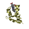
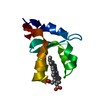
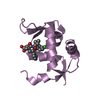
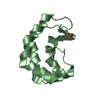
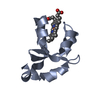
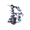
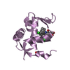
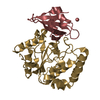
| Entry | Database: PDB / ID: 1b5m | ||||||
|---|---|---|---|---|---|---|---|
| Title | RAT OUTER MITOCHONDRIAL MEMBRANE CYTOCHROME B5 | ||||||
 Components Components | CYTOCHROME B5 | ||||||
 Keywords Keywords | ELECTRON TRANSPORT / CYTOCHROME / HEME | ||||||
| Function / homology |  Function and homology information Function and homology informationSphingolipid de novo biosynthesis / eNOS activation / Phase I - Functionalization of compounds / nitric-oxide synthase complex / quinol-cytochrome-c reductase activity / nitric oxide biosynthetic process / enzyme activator activity / mitochondrial outer membrane / heme binding / metal ion binding Similarity search - Function | ||||||
| Biological species |  | ||||||
| Method |  X-RAY DIFFRACTION / MOLECULAR REPLACEMENT / Resolution: 2.7 Å X-RAY DIFFRACTION / MOLECULAR REPLACEMENT / Resolution: 2.7 Å | ||||||
 Authors Authors | Rivera, M. / White, S.P. / Zhang, X. | ||||||
 Citation Citation | Journal: Biochemistry / Year: 1996 Title: 13C NMR spectroscopic and X-ray crystallographic study of the role played by mitochondrial cytochrome b5 heme propionates in the electrostatic binding to cytochrome c. Authors: Rodriguez-Maranon, M.J. / Qiu, F. / Stark, R.E. / White, S.P. / Zhang, X. / Foundling, S.I. / Rodriguez, V. / Schilling 3rd., C.L. / Bunce, R.A. / Rivera, M. | ||||||
| History |
|
- Structure visualization
| Structure viewer | Molecule: MolmilJmol/JSmol |
|---|
- Downloads & links
-Download
| PDBx/mmCIF format | 1b5m.cif.gz | 31.5 KB | Display | PDBx/mmCIF format |
|---|---|---|---|---|
| PDB format | pdb1b5m.ent.gz | 20.1 KB | Display | PDB format |
| PDBx/mmJSON format | 1b5m.json.gz | Tree view | PDBx/mmJSON format | |
| Others |  Other downloads Other downloads |
-Validation report
| Arichive directory | https://data.pdbj.org/pub/pdb/validation_reports/b5/1b5mftp://data.pdbj.org/pub/pdb/validation_reports/b5/1b5m | HTTPS FTP |
|---|
-Related structure data
| Related structure data |  3b5c S: Starting model for refinement |
|---|---|
| Similar structure data |


-Links
 PDBj
PDBj
- Assembly
| Deposited unit | 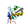
| ||||||||
|---|---|---|---|---|---|---|---|---|---|
| 1 |
| ||||||||
| Unit cell |
|
-Components
| #1: Protein | Mass: 9605.570 Da / Num. of mol.: 1 / Fragment: WATER SOLUBLE DOMAIN Source method: isolated from a genetically manipulated source Details: FROM RAT OUTER MITOCHONDRIAL MEMBRANE / Source: (gene. exp.)  |
|---|---|
| #2: Chemical | ChemComp-HEM /   Mass: 616.487 Da / Num. of mol.: 1 / Source method: obtained synthetically / Formula: C34H32FeN4O4 Mass: 616.487 Da / Num. of mol.: 1 / Source method: obtained synthetically / Formula: C34H32FeN4O4 |
-Experimental details
-Experiment
| Experiment | Method: X-RAY DIFFRACTION / Number of used crystals: 1 |
|---|
- Sample preparation
| Crystal | Density Matthews: 2.56 Å3/Da / Density % sol: 52.02 % | |||||||||||||||||||||||||
|---|---|---|---|---|---|---|---|---|---|---|---|---|---|---|---|---|---|---|---|---|---|---|---|---|---|---|
| Crystal grow | Method: vapor diffusion with slow evaporation / pH: 6.5 Details: CRYSTALS WERE OBTAINED BY VAPOR DIFFUSION TOGETHER WITH SLOW EVAPORATION USING 20% PEG 8000 IN 0.1M SODIUM CACODYLATE (PH = 6.5 WITH 0.2 M MAGNESIUM ACETATE., vapor diffusion with slow evaporation | |||||||||||||||||||||||||
| Crystal grow | *PLUS Method: vapor diffusion, hanging drop | |||||||||||||||||||||||||
| Components of the solutions | *PLUS
|
-Data collection
| Diffraction | Mean temperature: 298 K |
|---|---|
| Diffraction source | Source: ROTATING ANODE / Type: RIGAKU / Wavelength: 1.5418 |
| Detector | Type: SIEMENS / Detector: AREA DETECTOR / Date: Apr 18, 1996 |
| Radiation | Monochromator: DIAMOND C(111) / Monochromatic (M) / Laue (L): M / Scattering type: x-ray |
| Radiation wavelength | Wavelength: 1.5418 Å / Relative weight: 1 |
| Reflection | Resolution: 2.7→20 Å / Num. obs: 3123 / % possible obs: 96 % / Observed criterion σ(I): 0 / Redundancy: 2.5 % / Biso Wilson estimate: 36 Å2 / Rsym value: 0.049 / Net I/σ(I): 1.5 |
| Reflection shell | Resolution: 2.7→3.05 Å / Redundancy: 4 % / Mean I/σ(I) obs: 1 / Rsym value: 0.088 / % possible all: 85 |
| Reflection | *PLUS Rmerge(I) obs: 0.049 |
| Reflection shell | *PLUS % possible obs: 85 % / Rmerge(I) obs: 0.088 |
- Processing
| Software |
| ||||||||||||||||||||||||||||||||||||||||||||||||||
|---|---|---|---|---|---|---|---|---|---|---|---|---|---|---|---|---|---|---|---|---|---|---|---|---|---|---|---|---|---|---|---|---|---|---|---|---|---|---|---|---|---|---|---|---|---|---|---|---|---|---|---|
| Refinement | Method to determine structure: MOLECULAR REPLACEMENT Starting model: PDB ENTRY 3B5C 3b5c Resolution: 2.7→20 Å / Isotropic thermal model: TNT / σ(F): 0 / Stereochemistry target values: TNT
| ||||||||||||||||||||||||||||||||||||||||||||||||||
| Solvent computation | Solvent model: BSOL / Bsol: 99.7 Å2 / ksol: 0.79 e/Å3 | ||||||||||||||||||||||||||||||||||||||||||||||||||
| Refinement step | Cycle: LAST / Resolution: 2.7→20 Å
| ||||||||||||||||||||||||||||||||||||||||||||||||||
| Refine LS restraints |
| ||||||||||||||||||||||||||||||||||||||||||||||||||
| Software | *PLUS Name: TNT / Version: 5E / Classification: refinement | ||||||||||||||||||||||||||||||||||||||||||||||||||
| Refinement | *PLUS Rfactor obs: 0.22 | ||||||||||||||||||||||||||||||||||||||||||||||||||
| Solvent computation | *PLUS | ||||||||||||||||||||||||||||||||||||||||||||||||||
| Displacement parameters | *PLUS | ||||||||||||||||||||||||||||||||||||||||||||||||||
| Refine LS restraints | *PLUS Type: t_plane_restr / Dev ideal: 0.012 / Weight: 5 |
