 Movie
Movie Controller


+ Open data

- Basic information
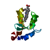
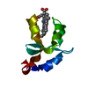
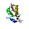
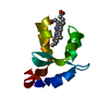
| Entry | Database: PDB / ID: 1lr6 | ||||||
|---|---|---|---|---|---|---|---|
| Title | Crystal structure of V45Y mutant of cytochrome b5 | ||||||
 Components Components | cytochrome b5 | ||||||
 Keywords Keywords | ELECTRON TRANSPORT / CYTOCHROME B5 / TRYPSIN-SOLUBILIZED FRAGMENT / MUTANT V45Y | ||||||
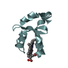
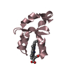
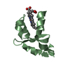
| Function / homology |  Function and homology information Function and homology informationVitamin C (ascorbate) metabolism / Insertion of tail-anchored proteins into the endoplasmic reticulum membrane / heme binding / endoplasmic reticulum membrane / metal ion binding Similarity search - Function | ||||||
| Biological species |  | ||||||
| Method |  X-RAY DIFFRACTION / FOURIER SYNTHESIS / Resolution: 1.9 Å X-RAY DIFFRACTION / FOURIER SYNTHESIS / Resolution: 1.9 Å | ||||||
 Authors Authors | Gan, J.-H. / Wu, J. / Wang, Z.-Q. / Wang, Y.-H. / Huang, Z.-X. / Xia, Z.-X. | ||||||
 Citation Citation | Journal: Acta Crystallogr.,Sect.D / Year: 2002 Title: Structures of V45E and V45Y mutants and structure comparison of a variety of cytochrome b5 mutants. Authors: Gan, J.H. / Wu, J. / Wang, Z.Q. / Wang, Y.H. / Huang, Z.X. / Xia, Z.X. | ||||||
| History |
|
- Structure visualization
| Structure viewer | Molecule: MolmilJmol/JSmol |
|---|
- Downloads & links
-Download
| PDBx/mmCIF format | 1lr6.cif.gz | 33.7 KB | Display | PDBx/mmCIF format |
|---|---|---|---|---|
| PDB format | pdb1lr6.ent.gz | 21.3 KB | Display | PDB format |
| PDBx/mmJSON format | 1lr6.json.gz | Tree view | PDBx/mmJSON format | |
| Others |  Other downloads Other downloads |
-Validation report
| Arichive directory | https://data.pdbj.org/pub/pdb/validation_reports/lr/1lr6ftp://data.pdbj.org/pub/pdb/validation_reports/lr/1lr6 | HTTPS FTP |
|---|
-Related structure data
| Related structure data | 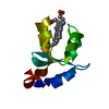 1lqxC 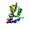 1ehbS S: Starting model for refinement C: citing same article ( |
|---|---|
| Similar structure data |


-Links
 PDBj
PDBj
- Assembly
| Deposited unit | 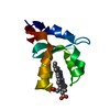
| ||||||||
|---|---|---|---|---|---|---|---|---|---|
| 1 |
| ||||||||
| Unit cell |
|
-Components
| #1: Protein | Mass: 9539.423 Da / Num. of mol.: 1 / Fragment: trypsin-solubilized fragment of cytochrome b5 / Mutation: V45Y Source method: isolated from a genetically manipulated source Source: (gene. exp.)  |
|---|---|
| #2: Chemical | ChemComp-HEM /   Mass: 616.487 Da / Num. of mol.: 1 / Source method: obtained synthetically / Formula: C34H32FeN4O4 Mass: 616.487 Da / Num. of mol.: 1 / Source method: obtained synthetically / Formula: C34H32FeN4O4 |
| #3: Water | ChemComp-HOH /  Mass: 18.015 Da / Num. of mol.: 90 / Source method: isolated from a natural source / Formula: H2O Mass: 18.015 Da / Num. of mol.: 90 / Source method: isolated from a natural source / Formula: H2O |
-Experimental details
-Experiment
| Experiment | Method: X-RAY DIFFRACTION / Number of used crystals: 1 |
|---|
- Sample preparation
| Crystal | Density Matthews: 2.68 Å3/Da / Density % sol: 54.07 % | ||||||||||||||||||
|---|---|---|---|---|---|---|---|---|---|---|---|---|---|---|---|---|---|---|---|
| Crystal grow | Temperature: 293 K / Method: vapor diffusion, hanging drop / pH: 7.5 Details: PHOSPHATE BUFFER, pH 7.5, VAPOR DIFFUSION, HANGING DROP, temperature 293.0K | ||||||||||||||||||
| Crystal grow | *PLUS Temperature: 293 K | ||||||||||||||||||
| Components of the solutions | *PLUS
|
-Data collection
| Diffraction | Mean temperature: 293 K |
|---|---|
| Diffraction source | Source: SEALED TUBE / Wavelength: 1.5418 Å |
| Detector | Type: MARRESEARCH / Detector: IMAGE PLATE / Date: Nov 10, 1999 |
| Radiation | Protocol: SINGLE WAVELENGTH / Monochromatic (M) / Laue (L): M / Scattering type: x-ray |
| Radiation wavelength | Wavelength: 1.5418 Å / Relative weight: 1 |
| Reflection | Resolution: 1.9→22.82 Å / Num. obs: 7563 / % possible obs: 93.5 % / Redundancy: 3.1 % / Biso Wilson estimate: 16 Å2 / Rmerge(I) obs: 0.062 |
| Reflection shell | Resolution: 1.9→1.94 Å / Redundancy: 2.3 % / Rmerge(I) obs: 0.194 / Mean I/σ(I) obs: 6.1 / % possible all: 76.3 |
| Reflection | *PLUS Highest resolution: 1.9 Å / Rmerge(I) obs: 0.062 |
| Reflection shell | *PLUS % possible obs: 76.3 % / Rmerge(I) obs: 0.194 |
- Processing
| Software |
| ||||||||||||||||||||||||||||||||||||||||||||||||||||||||||||
|---|---|---|---|---|---|---|---|---|---|---|---|---|---|---|---|---|---|---|---|---|---|---|---|---|---|---|---|---|---|---|---|---|---|---|---|---|---|---|---|---|---|---|---|---|---|---|---|---|---|---|---|---|---|---|---|---|---|---|---|---|---|
| Refinement | Method to determine structure: FOURIER SYNTHESIS Starting model: PDB ENTRY 1EHB Resolution: 1.9→22.82 Å / Rfactor Rfree error: 0.008 / Isotropic thermal model: RESTRAINED / Cross valid method: THROUGHOUT / σ(F): 0 / σ(I): 0
| ||||||||||||||||||||||||||||||||||||||||||||||||||||||||||||
| Solvent computation | Solvent model: FLAT MODEL / Bsol: 66.6757 Å2 / ksol: 0.402579 e/Å3 | ||||||||||||||||||||||||||||||||||||||||||||||||||||||||||||
| Displacement parameters | Biso mean: 29.1 Å2
| ||||||||||||||||||||||||||||||||||||||||||||||||||||||||||||
| Refine analyze | Luzzati coordinate error free: 0.27 Å / Luzzati sigma a free: 0.19 Å | ||||||||||||||||||||||||||||||||||||||||||||||||||||||||||||
| Refinement step | Cycle: LAST / Resolution: 1.9→22.82 Å
| ||||||||||||||||||||||||||||||||||||||||||||||||||||||||||||
| Refine LS restraints |
| ||||||||||||||||||||||||||||||||||||||||||||||||||||||||||||
| LS refinement shell | Resolution: 1.9→1.97 Å / Rfactor Rfree error: 0.035 / Total num. of bins used: 10
| ||||||||||||||||||||||||||||||||||||||||||||||||||||||||||||
| Xplor file |
| ||||||||||||||||||||||||||||||||||||||||||||||||||||||||||||
| Refinement | *PLUS Highest resolution: 1.9 Å / Rfactor all: 0.196 / Rfactor obs: 0.191 / Rfactor Rfree: 0.235 / Rfactor Rwork: 0.192 | ||||||||||||||||||||||||||||||||||||||||||||||||||||||||||||
| Solvent computation | *PLUS | ||||||||||||||||||||||||||||||||||||||||||||||||||||||||||||
| Displacement parameters | *PLUS | ||||||||||||||||||||||||||||||||||||||||||||||||||||||||||||
| Refine LS restraints | *PLUS
| ||||||||||||||||||||||||||||||||||||||||||||||||||||||||||||
| LS refinement shell | *PLUS Rfactor Rfree: 0.274 / Rfactor Rwork: 0.289 |
