 Movie
Movie Controller

[English] 日本語
 Yorodumi
- PDB-1m20: Crystal Structure of F35Y Mutant of Trypsin-solubilized Fragment ... -
+ Open data

- Basic information
| Entry | Database: PDB / ID: 1m20 | ||||||
|---|---|---|---|---|---|---|---|
| Title | Crystal Structure of F35Y Mutant of Trypsin-solubilized Fragment of Cytochrome b5 | ||||||
 Components Components | Cytochrome b5 | ||||||
 Keywords Keywords | ELECTRON TRANSPORT / cytochrome b5 / trypsin-cleaved fragment / mutant F35Y | ||||||
| Function / homology |  Function and homology information Function and homology informationVitamin C (ascorbate) metabolism / Insertion of tail-anchored proteins into the endoplasmic reticulum membrane / heme binding / endoplasmic reticulum membrane / metal ion binding Similarity search - Function | ||||||
| Biological species |  | ||||||
| Method |  X-RAY DIFFRACTION / FOURIER SYNTHESIS / Resolution: 1.8 Å X-RAY DIFFRACTION / FOURIER SYNTHESIS / Resolution: 1.8 Å | ||||||
 Authors Authors | Yao, P. / Wu, J. / Wang, Y.-H. / Sun, B.-Y. / Xia, Z.-X. / Huang, Z.-X. | ||||||
 Citation Citation | Journal: Eur.J.Biochem. / Year: 2002 Title: X-ray crystallography, CD and kinetic studies revealed the essence of the abnormal behaviors of the cytochrome b5 Phe35-->Tyr mutant. Authors: Yao, P. / Wu, J. / Wang, Y.H. / Sun, B.Y. / Xia, Z.X. / Huang, Z.X. | ||||||
| History |
|
- Structure visualization
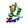
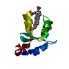
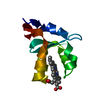
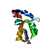
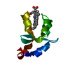
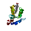
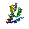
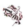
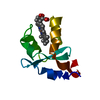
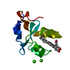
| Structure viewer | Molecule: MolmilJmol/JSmol |
|---|
- Downloads & links
-Download
| PDBx/mmCIF format | 1m20.cif.gz | 33.8 KB | Display | PDBx/mmCIF format |
|---|---|---|---|---|
| PDB format | pdb1m20.ent.gz | 21.4 KB | Display | PDB format |
| PDBx/mmJSON format | 1m20.json.gz | Tree view | PDBx/mmJSON format | |
| Others |  Other downloads Other downloads |
-Validation report
| Arichive directory | https://data.pdbj.org/pub/pdb/validation_reports/m2/1m20ftp://data.pdbj.org/pub/pdb/validation_reports/m2/1m20 | HTTPS FTP |
|---|
-Related structure data
| Related structure data |  1es1S S: Starting model for refinement |
|---|---|
| Similar structure data |


-Links
 PDBj
PDBj
- Assembly
| Deposited unit | 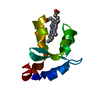
| ||||||||
|---|---|---|---|---|---|---|---|---|---|
| 1 |
| ||||||||
| Unit cell |
|
-Components
| #1: Protein | Mass: 9491.380 Da / Num. of mol.: 1 / Fragment: trypsin-solubilized fragment / Mutation: F35Y Source method: isolated from a genetically manipulated source Source: (gene. exp.)  |
|---|---|
| #2: Chemical | ChemComp-HEM /   Mass: 616.487 Da / Num. of mol.: 1 / Source method: obtained synthetically / Formula: C34H32FeN4O4 Mass: 616.487 Da / Num. of mol.: 1 / Source method: obtained synthetically / Formula: C34H32FeN4O4 |
| #3: Water | ChemComp-HOH /  Mass: 18.015 Da / Num. of mol.: 94 / Source method: isolated from a natural source / Formula: H2O Mass: 18.015 Da / Num. of mol.: 94 / Source method: isolated from a natural source / Formula: H2O |
-Experimental details
-Experiment
| Experiment | Method: X-RAY DIFFRACTION / Number of used crystals: 1 |
|---|
- Sample preparation
| Crystal | Density Matthews: 2.75 Å3/Da / Density % sol: 55.2 % | ||||||||||||||||||
|---|---|---|---|---|---|---|---|---|---|---|---|---|---|---|---|---|---|---|---|
| Crystal grow | Temperature: 293 K / Method: vapor diffusion, hanging drop / pH: 7.5 Details: phosphate buffer, pH 7.5, VAPOR DIFFUSION, HANGING DROP, temperature 293K | ||||||||||||||||||
| Crystal grow | *PLUS Temperature: 20 ℃ | ||||||||||||||||||
| Components of the solutions | *PLUS
|
-Data collection
| Diffraction | Mean temperature: 293 K |
|---|---|
| Diffraction source | Source: SEALED TUBE / Wavelength: 1.5418 Å |
| Detector | Type: MARRESEARCH / Detector: IMAGE PLATE |
| Radiation | Protocol: SINGLE WAVELENGTH / Monochromatic (M) / Laue (L): M / Scattering type: x-ray |
| Radiation wavelength | Wavelength: 1.5418 Å / Relative weight: 1 |
| Reflection | Resolution: 1.8→19.91 Å / Num. all: 9129 / Num. obs: 8384 / Biso Wilson estimate: 20.2 Å2 / Rsym value: 0.061 |
| Reflection shell | Resolution: 1.8→1.84 Å / Num. unique all: 511 / Rsym value: 0.315 |
| Reflection | *PLUS Highest resolution: 1.8 Å / Num. obs: 9129 / % possible obs: 94.3 % / Rmerge(I) obs: 0.061 |
| Reflection shell | *PLUS % possible obs: 79.7 % / Rmerge(I) obs: 0.315 / Mean I/σ(I) obs: 3.9 |
- Processing
| Software |
| ||||||||||||||||||||||||||||||||||||
|---|---|---|---|---|---|---|---|---|---|---|---|---|---|---|---|---|---|---|---|---|---|---|---|---|---|---|---|---|---|---|---|---|---|---|---|---|---|
| Refinement | Method to determine structure: FOURIER SYNTHESIS Starting model: PDB Entry 1ES1 Resolution: 1.8→19.91 Å / Rfactor Rfree error: 0.007 / Data cutoff high absF: 589664.97 / Data cutoff low absF: 0 / Isotropic thermal model: RESTRAINED / Cross valid method: THROUGHOUT / σ(F): 0
| ||||||||||||||||||||||||||||||||||||
| Solvent computation | Solvent model: FLAT MODEL / Bsol: 39.0714 Å2 / ksol: 0.351386 e/Å3 | ||||||||||||||||||||||||||||||||||||
| Displacement parameters | Biso mean: 26.4 Å2
| ||||||||||||||||||||||||||||||||||||
| Refine analyze |
| ||||||||||||||||||||||||||||||||||||
| Refinement step | Cycle: LAST / Resolution: 1.8→19.91 Å
| ||||||||||||||||||||||||||||||||||||
| Refine LS restraints |
| ||||||||||||||||||||||||||||||||||||
| LS refinement shell | Resolution: 1.8→1.86 Å / Rfactor Rfree error: 0.035 / Total num. of bins used: 10
| ||||||||||||||||||||||||||||||||||||
| Xplor file |
| ||||||||||||||||||||||||||||||||||||
| Refinement | *PLUS Rfactor obs: 0.193 / Rfactor Rfree: 0.238 / Rfactor Rwork: 0.192 | ||||||||||||||||||||||||||||||||||||
| Solvent computation | *PLUS | ||||||||||||||||||||||||||||||||||||
| Displacement parameters | *PLUS | ||||||||||||||||||||||||||||||||||||
| Refine LS restraints | *PLUS
| ||||||||||||||||||||||||||||||||||||
| LS refinement shell | *PLUS Rfactor Rfree: 0.302 / Rfactor Rwork: 0.306 |
