 Movie
Movie Controller

[English] 日本語
 Yorodumi
- PDB-1lj0: Structure of quintuple mutant of the rat outer mitocondrial cytoc... -
+ Open data

- Basic information
| Entry | Database: PDB / ID: 1lj0 | ||||||
|---|---|---|---|---|---|---|---|
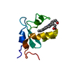
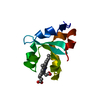
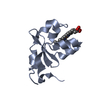
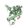
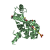
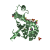
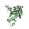
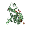
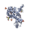
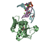
| Title | Structure of quintuple mutant of the rat outer mitocondrial cytochrome b5. | ||||||
 Components Components | Cytochrome B5 outer mitochondrial membrane isoform | ||||||
 Keywords Keywords | ELECTRON TRANSPORT / CYTOCHROME / HEME / protein engineering | ||||||
| Function / homology |  Function and homology information Function and homology informationSphingolipid de novo biosynthesis / eNOS activation / Phase I - Functionalization of compounds / nitric-oxide synthase complex / quinol-cytochrome-c reductase activity / nitric oxide biosynthetic process / enzyme activator activity / mitochondrial outer membrane / heme binding / metal ion binding Similarity search - Function | ||||||
| Biological species |  | ||||||
| Method |  X-RAY DIFFRACTION / FOURIER SYNTHESIS / Resolution: 2 Å X-RAY DIFFRACTION / FOURIER SYNTHESIS / Resolution: 2 Å | ||||||
 Authors Authors | Cowley, A.B. / Altuve, A. / Kuchment, O. / Terzyan, S. / Zhang, X.C. / Rivera, M. / Benson, D. | ||||||
 Citation Citation | Journal: Biochemistry / Year: 2002 Title: Toward engineering the stability and hemin binding properties of microsomal cytochromes b5 into rat outer mitochondrial cytochrome b5: Examining the influence of residues 25 and 71. Authors: Cowley, A.B. / Altuve, A. / Kuchment, O. / Terzyan, S. / Zhang, X.C. / Rivera, M. / Benson, D. | ||||||
| History |
|
- Structure visualization
| Structure viewer | Molecule: MolmilJmol/JSmol |
|---|
- Downloads & links
-Download
| PDBx/mmCIF format | 1lj0.cif.gz | 92.3 KB | Display | PDBx/mmCIF format |
|---|---|---|---|---|
| PDB format | pdb1lj0.ent.gz | 70.6 KB | Display | PDB format |
| PDBx/mmJSON format | 1lj0.json.gz | Tree view | PDBx/mmJSON format | |
| Others |  Other downloads Other downloads |
-Validation report
| Arichive directory | https://data.pdbj.org/pub/pdb/validation_reports/lj/1lj0ftp://data.pdbj.org/pub/pdb/validation_reports/lj/1lj0 | HTTPS FTP |
|---|
-Related structure data
| Related structure data | 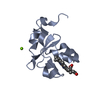 1iccS S: Starting model for refinement |
|---|---|
| Similar structure data |


-Links
 PDBj
PDBj
- Assembly
| Deposited unit | 
| ||||||||
|---|---|---|---|---|---|---|---|---|---|
| 1 | 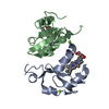
| ||||||||
| 2 | 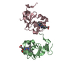
| ||||||||
| 3 | 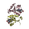
| ||||||||
| 4 | 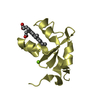
| ||||||||
| 5 | 
| ||||||||
| 6 | 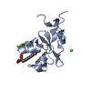
| ||||||||
| Unit cell |
|
-Components
| #1: Protein | Mass: 10424.323 Da / Num. of mol.: 4 / Fragment: water soluble domain / Mutation: A18S, I25L, I32L, L47R, L71S Source method: isolated from a genetically manipulated source Source: (gene. exp.)  #2: Chemical |   Mass: 24.305 Da / Num. of mol.: 3 / Source method: obtained synthetically / Formula: Mg Mass: 24.305 Da / Num. of mol.: 3 / Source method: obtained synthetically / Formula: Mg#3: Chemical | ChemComp-HEM /   Mass: 616.487 Da / Num. of mol.: 4 / Source method: obtained synthetically / Formula: C34H32FeN4O4 Mass: 616.487 Da / Num. of mol.: 4 / Source method: obtained synthetically / Formula: C34H32FeN4O4#4: Water | ChemComp-HOH / |  Mass: 18.015 Da / Num. of mol.: 191 / Source method: isolated from a natural source / Formula: H2O Mass: 18.015 Da / Num. of mol.: 191 / Source method: isolated from a natural source / Formula: H2O |
|---|
-Experimental details
-Experiment
| Experiment | Method: X-RAY DIFFRACTION / Number of used crystals: 1 |
|---|
- Sample preparation
| Crystal | Density Matthews: 2.03 Å3/Da / Density % sol: 35 % | ||||||||||||||||||||||||||||||
|---|---|---|---|---|---|---|---|---|---|---|---|---|---|---|---|---|---|---|---|---|---|---|---|---|---|---|---|---|---|---|---|
| Crystal grow | Temperature: 278 K / Method: vapor diffusion, hanging drop / pH: 6.5 Details: PEG 8000, MG ACETATE, pH 6.5, VAPOR DIFFUSION, HANGING DROP, temperature 278K | ||||||||||||||||||||||||||||||
| Crystal grow | *PLUS | ||||||||||||||||||||||||||||||
| Components of the solutions | *PLUS
|
-Data collection
| Diffraction | Mean temperature: 100 K |
|---|---|
| Diffraction source | Source: ROTATING ANODE / Type: RIGAKU RU300 / Wavelength: 1.54 Å |
| Detector | Type: MARRESEARCH / Detector: IMAGE PLATE / Date: Feb 8, 2002 / Details: Osmic multilayer mirrors |
| Radiation | Protocol: SINGLE WAVELENGTH / Monochromatic (M) / Laue (L): M / Scattering type: x-ray |
| Radiation wavelength | Wavelength: 1.54 Å / Relative weight: 1 |
| Reflection | Resolution: 2→50 Å / Num. obs: 23729 / % possible obs: 99.2 % / Observed criterion σ(I): -3 / Redundancy: 5 % / Biso Wilson estimate: 33.02 Å2 / Rmerge(I) obs: 0.08 / Net I/σ(I): 22 |
| Reflection shell | Resolution: 2→2.1 Å / Redundancy: 3.78 % / Rmerge(I) obs: 0.37 / Mean I/σ(I) obs: 3.2 / Num. unique all: 2215 / % possible all: 94.7 |
| Reflection shell | *PLUS % possible obs: 94.7 % |
- Processing
| Software |
| |||||||||||||||||||||||||
|---|---|---|---|---|---|---|---|---|---|---|---|---|---|---|---|---|---|---|---|---|---|---|---|---|---|---|
| Refinement | Method to determine structure: FOURIER SYNTHESIS Starting model: pdb entry 1ICC Resolution: 2→23 Å / Isotropic thermal model: Overall anisotropic B factor / Cross valid method: R free / σ(F): 0 / Stereochemistry target values: Engh & Huber / Details: USED MAXIMUM LIKELIHOOD TARGET FOR AMPLITUDES
| |||||||||||||||||||||||||
| Solvent computation | Bsol: 49.18 Å2 / ksol: 0.37 e/Å3 | |||||||||||||||||||||||||
| Displacement parameters | Biso mean: 34.5 Å2
| |||||||||||||||||||||||||
| Refinement step | Cycle: LAST / Resolution: 2→23 Å
| |||||||||||||||||||||||||
| Refine LS restraints |
| |||||||||||||||||||||||||
| Refinement | *PLUS Num. reflection obs: 20088 / Rfactor Rwork: 0.21 | |||||||||||||||||||||||||
| Solvent computation | *PLUS | |||||||||||||||||||||||||
| Displacement parameters | *PLUS |
