 Movie
Movie Controller


+ Open data

- Basic information
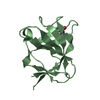
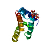
| Entry | Database: PDB / ID: 1cyo | |||||||||
|---|---|---|---|---|---|---|---|---|---|---|
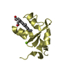
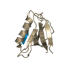
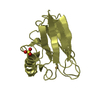
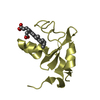
| Title | BOVINE CYTOCHROME B(5) | |||||||||
 Components Components | CYTOCHROME B5 | |||||||||
 Keywords Keywords | ELECTRON TRANSPORT | |||||||||
| Function / homology |  Function and homology information Function and homology informationVitamin C (ascorbate) metabolism / Insertion of tail-anchored proteins into the endoplasmic reticulum membrane / heme binding / endoplasmic reticulum membrane / metal ion binding Similarity search - Function | |||||||||
| Biological species |  | |||||||||
| Method |  X-RAY DIFFRACTION / Resolution: 1.5 Å X-RAY DIFFRACTION / Resolution: 1.5 Å | |||||||||
 Authors Authors | Durley, R.C.E. / Mathews, F.S. | |||||||||
 Citation Citation | Journal: Acta Crystallogr.,Sect.D / Year: 1996 Title: Refinement and structural analysis of bovine cytochrome b5 at 1.5 A resolution. Authors: Durley, R.C. / Mathews, F.S. #1: Journal: The Enzymes of Biological Membranes. 2Nd Ed. V.4: Bioenergetics of Electron and Proton TransportYear: 1985 Title: Cytochrome B5 and Cytochrome B5 Reductase from a Chemical and X-Ray Diffraction Viewpoint Authors: Mathews, F.S. / Czerwinski, E.W. #2: Journal: The Porphyrins V.7: Biochemistry, Pt.B / Year: 1979Title: The X-Ray Crystallographic Structure of Calf Liver Cytochrome B5 Authors: Mathews, F.S. / Czerwinski, E.W. / Argos, P. #3: Journal: Cold Spring Harbor Symp.Quant.Biol. / Year: 1972Title: The Structure of Cytochrome B5 at 2.0 Angstroms Resolution Authors: Mathews, F.S. / Argos, P. / Levine, M. | |||||||||
| History |
|
- Structure visualization
| Structure viewer | Molecule: MolmilJmol/JSmol |
|---|
- Downloads & links
-Download
| PDBx/mmCIF format | 1cyo.cif.gz | 34.4 KB | Display | PDBx/mmCIF format |
|---|---|---|---|---|
| PDB format | pdb1cyo.ent.gz | 22.8 KB | Display | PDB format |
| PDBx/mmJSON format | 1cyo.json.gz | Tree view | PDBx/mmJSON format | |
| Others |  Other downloads Other downloads |
-Validation report
| Arichive directory | https://data.pdbj.org/pub/pdb/validation_reports/cy/1cyoftp://data.pdbj.org/pub/pdb/validation_reports/cy/1cyo | HTTPS FTP |
|---|
-Related structure data
| Similar structure data |
|---|


-Links
 PDBj
PDBj
- Assembly
| Deposited unit | 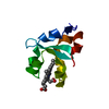
| ||||||||
|---|---|---|---|---|---|---|---|---|---|
| 1 |
| ||||||||
| Unit cell |
| ||||||||
| Atom site foot note | 1: ALTERNATE POSITIONS WERE REFINED FOR THE SIDE-CHAINS OF RESIDUES GLU 48, VAL 61, GLU 69, AND ILE 75. |
-Components
| #1: Protein | Mass: 10651.721 Da / Num. of mol.: 1 Source method: isolated from a genetically manipulated source Source: (gene. exp.) |
|---|---|
| #2: Chemical | ChemComp-HEM /   Mass: 616.487 Da / Num. of mol.: 1 / Source method: obtained synthetically / Formula: C34H32FeN4O4 Mass: 616.487 Da / Num. of mol.: 1 / Source method: obtained synthetically / Formula: C34H32FeN4O4 |
| #3: Water | ChemComp-HOH /  Mass: 18.015 Da / Num. of mol.: 117 / Source method: isolated from a natural source / Formula: H2O Mass: 18.015 Da / Num. of mol.: 117 / Source method: isolated from a natural source / Formula: H2O |
| Compound details | THE PROTEIN WAS SOLUBILIZE |
-Experimental details
-Experiment
| Experiment | Method: X-RAY DIFFRACTION |
|---|
- Sample preparation
| Crystal | Density Matthews: 2.09 Å3/Da / Density % sol: 41.02 % | ||||||||||||||||||||
|---|---|---|---|---|---|---|---|---|---|---|---|---|---|---|---|---|---|---|---|---|---|
| Crystal grow | *PLUS pH: 7.1 / Method: vapor diffusion, hanging drop | ||||||||||||||||||||
| Components of the solutions | *PLUS
|
-Data collection
| Reflection | *PLUS Highest resolution: 1.5 Å / Lowest resolution: 2.2 Å / Num. obs: 9843 / % possible obs: 99.2 % / Num. measured all: 13356 |
|---|---|
| Reflection shell | *PLUS Highest resolution: 1.5 Å / Lowest resolution: 1.6 Å / % possible obs: 16.5 % |
- Processing
| Software |
| ||||||||||||||||||||||||||||||||||||||||||||||||||||||||||||
|---|---|---|---|---|---|---|---|---|---|---|---|---|---|---|---|---|---|---|---|---|---|---|---|---|---|---|---|---|---|---|---|---|---|---|---|---|---|---|---|---|---|---|---|---|---|---|---|---|---|---|---|---|---|---|---|---|---|---|---|---|---|
| Refinement | Resolution: 1.5→10 Å /
| ||||||||||||||||||||||||||||||||||||||||||||||||||||||||||||
| Refinement step | Cycle: LAST / Resolution: 1.5→10 Å
| ||||||||||||||||||||||||||||||||||||||||||||||||||||||||||||
| Refine LS restraints |
| ||||||||||||||||||||||||||||||||||||||||||||||||||||||||||||
| Refinement | *PLUS Rfactor Rwork: 0.164 | ||||||||||||||||||||||||||||||||||||||||||||||||||||||||||||
| Solvent computation | *PLUS | ||||||||||||||||||||||||||||||||||||||||||||||||||||||||||||
| Displacement parameters | *PLUS | ||||||||||||||||||||||||||||||||||||||||||||||||||||||||||||
| Refine LS restraints | *PLUS Type: x_bond_d / Dev ideal: 0.009 |
