 Movie
Movie Controller

[English] 日本語
 Yorodumi
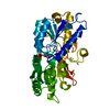
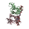
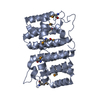
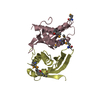
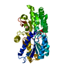
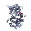
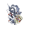
- PDB-2i3g: Crystal structure of N-Acetyl-gamma-Glutamyl-Phosphate Reductase ... -
+ Open data

- Basic information
| Entry | Database: PDB / ID: 2i3g | ||||||
|---|---|---|---|---|---|---|---|
| Title | Crystal structure of N-Acetyl-gamma-Glutamyl-Phosphate Reductase (Rv1652) from Mycobacterium tuberculosis in complex with NADP+. | ||||||
 Components Components | N-acetyl-gamma-glutamyl-phosphate reductase | ||||||
 Keywords Keywords | OXIDOREDUCTASE / dimer interface beta sandwich / dimer / Rossmann fold / Structural Genomics / Mycobacterium Tuberculosis Structural Proteomics Project / XMTB / TB Structural Genomics Consortium / TBSGC | ||||||
| Function / homology |  Function and homology information Function and homology informationN-acetyl-gamma-glutamyl-phosphate reductase / N-acetyl-gamma-glutamyl-phosphate reductase activity / L-arginine biosynthetic process / NADP+ binding / NAD binding / protein dimerization activity / cytoplasm Similarity search - Function | ||||||
| Biological species |   Mycobacterium tuberculosis (bacteria) Mycobacterium tuberculosis (bacteria) | ||||||
| Method |  X-RAY DIFFRACTION / SYNCHROTRON / MOLECULAR REPLACEMENT / Resolution: 1.85 Å X-RAY DIFFRACTION / SYNCHROTRON / MOLECULAR REPLACEMENT / Resolution: 1.85 Å | ||||||
 Authors Authors | Cherney, L.T. / Cherney, M.M. / Garen, C.R. / Moraidin, F. / James, M.N.G. / Mycobacterium Tuberculosis Structural Proteomics Project (XMTB) / TB Structural Genomics Consortium (TBSGC) | ||||||
 Citation Citation | Journal: J.Mol.Biol. / Year: 2007 Title: Crystal Structure of N-acetyl-gamma-glutamyl-phosphate Reductase from Mycobacterium tuberculosis in Complex with NADP(+). Authors: Cherney, L.T. / Cherney, M.M. / Garen, C.R. / Niu, C. / Moradian, F. / James, M.N.G. | ||||||
| History |
| ||||||
| Remark 300 | BIOMOLECULE: 1, 2 THIS ENTRY CONTAINS THE CRYSTALLOGRAPHIC ASYMMETRIC UNIT WHICH CONSISTS OF 2 ...BIOMOLECULE: 1, 2 THIS ENTRY CONTAINS THE CRYSTALLOGRAPHIC ASYMMETRIC UNIT WHICH CONSISTS OF 2 CHAIN(S). SEE REMARK 350 FOR INFORMATION ON GENERATING THE BIOLOGICAL MOLECULE(S). THE BIOLOGICAL UNIT HAS BEEN CORRECTED BY TRANSLATING CHAIN A WITH SYMMETRY OPERATOR -X,Y,-Z. |
- Structure visualization
| Structure viewer | Molecule: MolmilJmol/JSmol |
|---|
- Downloads & links
-Download
| PDBx/mmCIF format | 2i3g.cif.gz | 157.3 KB | Display | PDBx/mmCIF format |
|---|---|---|---|---|
| PDB format | pdb2i3g.ent.gz | 122.3 KB | Display | PDB format |
| PDBx/mmJSON format | 2i3g.json.gz | Tree view | PDBx/mmJSON format | |
| Others |  Other downloads Other downloads |
-Validation report
| Arichive directory | https://data.pdbj.org/pub/pdb/validation_reports/i3/2i3gftp://data.pdbj.org/pub/pdb/validation_reports/i3/2i3g | HTTPS FTP |
|---|
-Related structure data
| Related structure data |  2i3aC  2nqtC  1vknS S: Starting model for refinement C: citing same article ( |
|---|---|
| Similar structure data | |
| Other databases |


-Links
 PDBj
PDBj- Assembly
| Deposited unit | 
| ||||||||
|---|---|---|---|---|---|---|---|---|---|
| 1 | 
| ||||||||
| 2 | 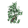
| ||||||||
| 3 | 
| ||||||||
| Unit cell |
| ||||||||
| Details | The biologycal assembly is a dimer. There are two half-dimers in the asymmetric unit. |
-Components
| #1: Protein | Mass: 36341.176 Da / Num. of mol.: 2 Source method: isolated from a genetically manipulated source Source: (gene. exp.) Mycobacterium tuberculosis (bacteria) / Strain: H37Rv / Gene: argC / Plasmid: pDEST15-1652 / Production host: References: UniProt: P63562, UniProt: P9WPZ9*PLUS, N-acetyl-gamma-glutamyl-phosphate reductase #2: Chemical |   Mass: 743.405 Da / Num. of mol.: 2 / Source method: obtained synthetically / Formula: C21H28N7O17P3 Mass: 743.405 Da / Num. of mol.: 2 / Source method: obtained synthetically / Formula: C21H28N7O17P3#3: Chemical |   Mass: 209.240 Da / Num. of mol.: 2 / Source method: obtained synthetically / Formula: C8H19NO5 / Comment: pH buffer*YM Mass: 209.240 Da / Num. of mol.: 2 / Source method: obtained synthetically / Formula: C8H19NO5 / Comment: pH buffer*YM#4: Water | ChemComp-HOH / |  Mass: 18.015 Da / Num. of mol.: 688 / Source method: isolated from a natural source / Formula: H2O Mass: 18.015 Da / Num. of mol.: 688 / Source method: isolated from a natural source / Formula: H2O |
|---|
-Experimental details
-Experiment
| Experiment | Method: X-RAY DIFFRACTION / Number of used crystals: 1 |
|---|
- Sample preparation
| Crystal | Density Matthews: 2.66 Å3/Da / Density % sol: 53.72 % |
|---|---|
| Crystal grow | Temperature: 295 K / Method: vapor diffusion, sitting drop / pH: 5.5 Details: 17% PEG 10000, 0.1 M Bis-Tris pH 5.5, 0.1 M Ammonium Acetate, VAPOR DIFFUSION, SITTING DROP, temperature 295K |
-Data collection
| Diffraction | Mean temperature: 100 K |
|---|---|
| Diffraction source | Source: SYNCHROTRON / Site: ALS  / Beamline: 8.3.1 / Wavelength: 1.11587 Å / Beamline: 8.3.1 / Wavelength: 1.11587 Å |
| Detector | Type: ADSC QUANTUM 210 / Detector: CCD / Date: Apr 14, 2006 |
| Radiation | Monochromator: single wavelength / Protocol: SINGLE WAVELENGTH / Monochromatic (M) / Laue (L): M / Scattering type: x-ray |
| Radiation wavelength | Wavelength: 1.11587 Å / Relative weight: 1 |
| Reflection | Resolution: 1.85→50 Å / Num. all: 65308 / Num. obs: 59626 / % possible obs: 91.3 % / Observed criterion σ(F): 0 / Observed criterion σ(I): 0 |
| Reflection shell | Resolution: 1.85→1.92 Å / % possible all: 88.2 |
- Processing
| Software |
| ||||||||||||||||||||||||||||||||||||||||||||||||||||||||||||||||||||||||||||||||||||||||||
|---|---|---|---|---|---|---|---|---|---|---|---|---|---|---|---|---|---|---|---|---|---|---|---|---|---|---|---|---|---|---|---|---|---|---|---|---|---|---|---|---|---|---|---|---|---|---|---|---|---|---|---|---|---|---|---|---|---|---|---|---|---|---|---|---|---|---|---|---|---|---|---|---|---|---|---|---|---|---|---|---|---|---|---|---|---|---|---|---|---|---|---|
| Refinement | Method to determine structure: MOLECULAR REPLACEMENT Starting model: PDB entry 1vkn Resolution: 1.85→50 Å / Cor.coef. Fo:Fc: 0.957 / Cor.coef. Fo:Fc free: 0.93 / SU B: 2.637 / SU ML: 0.08 / Cross valid method: THROUGHOUT / σ(F): 0 / ESU R: 0.132 / ESU R Free: 0.128 / Stereochemistry target values: MAXIMUM LIKELIHOOD / Details: HYDROGENS HAVE BEEN ADDED IN THE RIDING POSITIONS
| ||||||||||||||||||||||||||||||||||||||||||||||||||||||||||||||||||||||||||||||||||||||||||
| Solvent computation | Ion probe radii: 0.8 Å / Shrinkage radii: 0.8 Å / VDW probe radii: 1.4 Å / Solvent model: MASK | ||||||||||||||||||||||||||||||||||||||||||||||||||||||||||||||||||||||||||||||||||||||||||
| Displacement parameters | Biso mean: 14.308 Å2
| ||||||||||||||||||||||||||||||||||||||||||||||||||||||||||||||||||||||||||||||||||||||||||
| Refinement step | Cycle: LAST / Resolution: 1.85→50 Å
| ||||||||||||||||||||||||||||||||||||||||||||||||||||||||||||||||||||||||||||||||||||||||||
| Refine LS restraints |
| ||||||||||||||||||||||||||||||||||||||||||||||||||||||||||||||||||||||||||||||||||||||||||
| LS refinement shell | Resolution: 1.85→1.894 Å / Total num. of bins used: 20
|
