 Movie
Movie Controller

[English] 日本語
 Yorodumi
- PDB-2hle: Structural and biophysical characterization of the EPHB4-EPHRINB2... -
+ Open data

- Basic information
| Entry | Database: PDB / ID: 2hle | ||||||
|---|---|---|---|---|---|---|---|
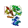
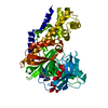
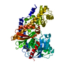
| Title | Structural and biophysical characterization of the EPHB4-EPHRINB2 protein protein interaction and receptor specificity. | ||||||
 Components Components |
| ||||||
 Keywords Keywords | TRANSFERASE/TRANSFERASE RECEPTOR / Protein-protein interaction / Receptor tryosine kinase / Bi-directional cell signaling / TRANSFERASE-TRANSFERASE RECEPTOR COMPLEX / Structural Genomics / PSI-2 / Protein Structure Initiative / Accelerated Technologies Center for Gene to 3D Structure / ATCG3D | ||||||
| Function / homology |  Function and homology information Function and homology informationpositive regulation of aorta morphogenesis / nephric duct morphogenesis / venous blood vessel morphogenesis / positive regulation of leukocyte adhesion to arterial endothelial cell / presynapse assembly / lymph vessel development / ephrin receptor activity / regulation of chemotaxis / positive regulation of cardiac muscle cell differentiation / cell migration involved in sprouting angiogenesis ...positive regulation of aorta morphogenesis / nephric duct morphogenesis / venous blood vessel morphogenesis / positive regulation of leukocyte adhesion to arterial endothelial cell / presynapse assembly / lymph vessel development / ephrin receptor activity / regulation of chemotaxis / positive regulation of cardiac muscle cell differentiation / cell migration involved in sprouting angiogenesis / adherens junction organization / blood vessel morphogenesis / EPH-Ephrin signaling / Ephrin signaling / regulation of postsynaptic neurotransmitter receptor internalization / keratinocyte proliferation / negative regulation of keratinocyte proliferation / EPH-ephrin mediated repulsion of cells / ephrin receptor signaling pathway / anatomical structure morphogenesis / regulation of postsynaptic membrane neurotransmitter receptor levels / heart morphogenesis / ephrin receptor binding / T cell costimulation / EPHB-mediated forward signaling / transmembrane receptor protein tyrosine kinase activity / axon guidance / animal organ morphogenesis / adherens junction / receptor protein-tyrosine kinase / postsynaptic density membrane / Schaffer collateral - CA1 synapse / negative regulation of neuron projection development / cellular response to lipopolysaccharide / virus receptor activity / angiogenesis / presynaptic membrane / cell adhesion / signaling receptor complex / receptor ligand activity / focal adhesion / positive regulation of cell population proliferation / dendrite / glutamatergic synapse / extracellular exosome / extracellular region / ATP binding / plasma membrane / cytosol Similarity search - Function | ||||||
| Biological species |  Homo sapiens (human) Homo sapiens (human) | ||||||
| Method |  X-RAY DIFFRACTION / SYNCHROTRON / MOLECULAR REPLACEMENT / Resolution: 2.05 Å X-RAY DIFFRACTION / SYNCHROTRON / MOLECULAR REPLACEMENT / Resolution: 2.05 Å | ||||||
 Authors Authors | Chrencik, J.E. / Brooun, A. / Kuhn, P. / Accelerated Technologies Center for Gene to 3D Structure (ATCG3D) | ||||||
 Citation Citation | Journal: J.Biol.Chem. / Year: 2006 Title: Structural and Biophysical Characterization of the EphB4-EphrinB2 Protein-Protein Interaction and Receptor Specificity. Authors: Chrencik, J.E. / Brooun, A. / Kraus, M.L. / Recht, M.I. / Kolatkar, A.R. / Han, G.W. / Seifert, J.M. / Widmer, H. / Auer, M. / Kuhn, P. | ||||||
| History |
|
- Structure visualization
| Structure viewer | Molecule: MolmilJmol/JSmol |
|---|
- Downloads & links
-Download
| PDBx/mmCIF format | 2hle.cif.gz | 80 KB | Display | PDBx/mmCIF format |
|---|---|---|---|---|
| PDB format | pdb2hle.ent.gz | 59.1 KB | Display | PDB format |
| PDBx/mmJSON format | 2hle.json.gz | Tree view | PDBx/mmJSON format | |
| Others |  Other downloads Other downloads |
-Validation report
| Arichive directory | https://data.pdbj.org/pub/pdb/validation_reports/hl/2hleftp://data.pdbj.org/pub/pdb/validation_reports/hl/2hle | HTTPS FTP |
|---|
-Related structure data
| Related structure data |  1kgyS S: Starting model for refinement |
|---|---|
| Similar structure data | |
| Other databases |


-Links
 PDBj
PDBj


- Assembly
| Deposited unit | 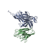
| ||||||||
|---|---|---|---|---|---|---|---|---|---|
| 1 |
| ||||||||
| Unit cell |
|
-Components
| #1: Protein | Mass: 21354.230 Da / Num. of mol.: 1 / Fragment: Ligand Binding Domain Source method: isolated from a genetically manipulated source Source: (gene. exp.) Homo sapiens (human) / Cell line: Hi5 / Gene: EPHB4, HTK / Production host:   Spodoptera frugiperda (fall armyworm) Spodoptera frugiperda (fall armyworm)References: UniProt: P54760, receptor protein-tyrosine kinase |
|---|---|
| #2: Protein | Mass: 15787.061 Da / Num. of mol.: 1 / Fragment: extracellular domain Source method: isolated from a genetically manipulated source Source: (gene. exp.) Homo sapiens (human) / Cell line: Hi5 / Gene: EFNB2, EPLG5, HTKL, LERK5 / Production host: Spodoptera frugiperda (fall armyworm) / References: UniProt: P52799 |
| #3: Water | ChemComp-HOH /  Mass: 18.015 Da / Num. of mol.: 79 / Source method: isolated from a natural source / Formula: H2O Mass: 18.015 Da / Num. of mol.: 79 / Source method: isolated from a natural source / Formula: H2O |
| Has protein modification | Y |
-Experimental details
-Experiment
| Experiment | Method: X-RAY DIFFRACTION / Number of used crystals: 1 |
|---|
- Sample preparation
| Crystal | Density Matthews: 2.25 Å3/Da / Density % sol: 45.42 % |
|---|---|
| Crystal grow | Temperature: 293 K / Method: vapor diffusion, sitting drop / pH: 7.8 Details: 2.2 M Ammonium sulfate, 100 mM Tris, pH 7.8, VAPOR DIFFUSION, SITTING DROP, temperature 293K |
-Data collection
| Diffraction | Mean temperature: 100 K |
|---|---|
| Diffraction source | Source: SYNCHROTRON / Site: APS  / Beamline: 22-ID / Wavelength: 1 Å / Beamline: 22-ID / Wavelength: 1 Å |
| Detector | Type: MAR CCD 165 mm / Detector: CCD / Date: Dec 1, 2005 |
| Radiation | Monochromator: Si 111 / Protocol: SINGLE WAVELENGTH / Monochromatic (M) / Laue (L): M / Scattering type: x-ray |
| Radiation wavelength | Wavelength: 1 Å / Relative weight: 1 |
| Reflection | Resolution: 2.05→20 Å / Num. obs: 19785 / % possible obs: 99.63 % / Observed criterion σ(F): 1 / Observed criterion σ(I): 1 |
| Reflection shell | Resolution: 2.05→2.103 Å / % possible all: 99.93 |
- Processing
| Software |
| |||||||||||||||||||||||||||||||||||||||||||||||||||||||||||||||||||||||||||||||||||||||||||||||||||||||||||||||||||||||||||||||||||||||||||||||||||||||||||||||||||||||||||||||
|---|---|---|---|---|---|---|---|---|---|---|---|---|---|---|---|---|---|---|---|---|---|---|---|---|---|---|---|---|---|---|---|---|---|---|---|---|---|---|---|---|---|---|---|---|---|---|---|---|---|---|---|---|---|---|---|---|---|---|---|---|---|---|---|---|---|---|---|---|---|---|---|---|---|---|---|---|---|---|---|---|---|---|---|---|---|---|---|---|---|---|---|---|---|---|---|---|---|---|---|---|---|---|---|---|---|---|---|---|---|---|---|---|---|---|---|---|---|---|---|---|---|---|---|---|---|---|---|---|---|---|---|---|---|---|---|---|---|---|---|---|---|---|---|---|---|---|---|---|---|---|---|---|---|---|---|---|---|---|---|---|---|---|---|---|---|---|---|---|---|---|---|---|---|---|---|---|
| Refinement | Method to determine structure: MOLECULAR REPLACEMENT Starting model: PDB ENTRY 1KGY Resolution: 2.05→20 Å / Cor.coef. Fo:Fc: 0.944 / Cor.coef. Fo:Fc free: 0.902 / SU B: 16.808 / SU ML: 0.225 / TLS residual ADP flag: LIKELY RESIDUAL / Cross valid method: THROUGHOUT / σ(F): 1 / ESU R: 0.245 / ESU R Free: 0.222 / Stereochemistry target values: MAXIMUM LIKELIHOOD Details: HYDROGENS HAVE BEEN ADDED IN THE RIDING POSITIONS.RESIDUES 74, 96-99 IN B MOLECULE WERE NOT VISIBLE IN THE ELECTRON DENSITY MAPS.
| |||||||||||||||||||||||||||||||||||||||||||||||||||||||||||||||||||||||||||||||||||||||||||||||||||||||||||||||||||||||||||||||||||||||||||||||||||||||||||||||||||||||||||||||
| Solvent computation | Ion probe radii: 0.8 Å / Shrinkage radii: 0.8 Å / VDW probe radii: 1.2 Å / Solvent model: MASK | |||||||||||||||||||||||||||||||||||||||||||||||||||||||||||||||||||||||||||||||||||||||||||||||||||||||||||||||||||||||||||||||||||||||||||||||||||||||||||||||||||||||||||||||
| Displacement parameters | Biso mean: 45.738 Å2
| |||||||||||||||||||||||||||||||||||||||||||||||||||||||||||||||||||||||||||||||||||||||||||||||||||||||||||||||||||||||||||||||||||||||||||||||||||||||||||||||||||||||||||||||
| Refinement step | Cycle: LAST / Resolution: 2.05→20 Å
| |||||||||||||||||||||||||||||||||||||||||||||||||||||||||||||||||||||||||||||||||||||||||||||||||||||||||||||||||||||||||||||||||||||||||||||||||||||||||||||||||||||||||||||||
| Refine LS restraints |
| |||||||||||||||||||||||||||||||||||||||||||||||||||||||||||||||||||||||||||||||||||||||||||||||||||||||||||||||||||||||||||||||||||||||||||||||||||||||||||||||||||||||||||||||
| LS refinement shell | Resolution: 2.05→2.103 Å / Total num. of bins used: 20
| |||||||||||||||||||||||||||||||||||||||||||||||||||||||||||||||||||||||||||||||||||||||||||||||||||||||||||||||||||||||||||||||||||||||||||||||||||||||||||||||||||||||||||||||
| Refinement TLS params. | Method: refined / Refine-ID: X-RAY DIFFRACTION
| |||||||||||||||||||||||||||||||||||||||||||||||||||||||||||||||||||||||||||||||||||||||||||||||||||||||||||||||||||||||||||||||||||||||||||||||||||||||||||||||||||||||||||||||
| Refinement TLS group |
|
