[English] 日本語
Yorodumi
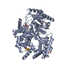
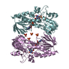
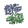
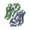
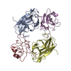
- PDB-1kgy: Crystal Structure of the EphB2-ephrinB2 complex -

+
Open data


ID or keywords:

Loading...

-
Basic information

Entry
Database: PDB / ID: 1kgy
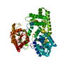
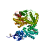
TitleCrystal Structure of the EphB2-ephrinB2 complex
Components
  • EPHRIN TYPE-B RECEPTOR 2
  • EPHRIN-B2
KeywordsTRANSFERASE/TRANSFERASE RECEPTOR / Developmental protein / TRANSFERASE-TRANSFERASE RECEPTOR COMPLEX
Function / homology
Function and homology information


regulation of T-helper 17 type immune response / nephric duct morphogenesis / positive regulation of aorta morphogenesis / Ephrin signaling / venous blood vessel morphogenesis / EPH-Ephrin signaling / urogenital system development / hindbrain tangential cell migration / negative regulation of glutamate receptor signaling pathway / optic nerve morphogenesis ...regulation of T-helper 17 type immune response / nephric duct morphogenesis / positive regulation of aorta morphogenesis / Ephrin signaling / venous blood vessel morphogenesis / EPH-Ephrin signaling / urogenital system development / hindbrain tangential cell migration / negative regulation of glutamate receptor signaling pathway / optic nerve morphogenesis / vesicle-mediated intercellular transport / EPH-ephrin mediated repulsion of cells / central nervous system projection neuron axonogenesis / neuron projection retraction / tight junction assembly / EPHB-mediated forward signaling / regulation of body fluid levels / postsynaptic membrane assembly / axon guidance receptor activity / dendritic spine development / transmembrane-ephrin receptor activity / positive regulation of leukocyte adhesion to arterial endothelial cell / lymph vessel development / negative regulation of axonogenesis / corpus callosum development / presynapse assembly / camera-type eye morphogenesis / positive regulation of long-term neuronal synaptic plasticity / retinal ganglion cell axon guidance / regulation of filopodium assembly / transmembrane receptor protein tyrosine kinase activator activity / ephrin receptor activity / adherens junction organization / positive regulation of protein localization to cell surface / positive regulation of cardiac muscle cell differentiation / regulation of chemotaxis / positive regulation of dendritic spine morphogenesis / dendritic spine morphogenesis / regulation of autophagosome assembly / negative regulation of cell adhesion / cell migration involved in sprouting angiogenesis / positive regulation of synaptic plasticity / blood vessel morphogenesis / negative regulation of Ras protein signal transduction / commissural neuron axon guidance / negative regulation of keratinocyte proliferation / axonal fasciculation / phosphorylation / positive regulation of synapse assembly / inner ear morphogenesis / regulation of behavioral fear response / roof of mouth development / regulation of receptor signaling pathway via JAK-STAT / regulation of axonogenesis / regulation of postsynaptic neurotransmitter receptor internalization / positive regulation of immunoglobulin production / regulation of blood coagulation / regulation of synapse assembly / positive regulation of glutamate receptor signaling pathway / regulation of neuronal synaptic plasticity / B cell activation / ephrin receptor signaling pathway / T cell costimulation / regulation of postsynaptic membrane neurotransmitter receptor levels / ephrin receptor binding / positive regulation of B cell proliferation / neuron projection maintenance / negative regulation of cytokine production involved in inflammatory response / peptidyl-tyrosine phosphorylation / animal organ morphogenesis / axon guidance / learning / cell surface receptor protein tyrosine kinase signaling pathway / hippocampal mossy fiber to CA3 synapse / positive regulation of long-term synaptic potentiation / positive regulation of protein localization to plasma membrane / negative regulation of neuron projection development / adherens junction / cell morphogenesis / negative regulation of ERK1 and ERK2 cascade / receptor protein-tyrosine kinase / postsynaptic density membrane / Schaffer collateral - CA1 synapse / cellular response to amyloid-beta / positive regulation of tumor necrosis factor production / amyloid-beta binding / cellular response to lipopolysaccharide / signaling receptor activity / angiogenesis / protein tyrosine kinase activity / presynaptic membrane / dendritic spine / learning or memory / protein phosphorylation / cell adhesion / postsynaptic membrane / postsynapse / positive regulation of cell migration / receptor ligand activity / signaling receptor binding
Similarity search - Function
Ephrin type-B receptor 2, ligand binding domain / Ephrin-B ectodomain / Ephrin receptor-binding domain / Ephrin, conserved site / Ephrin / Ephrin / Ephrin receptor-binding (ephrin RBD) domain signature. / Ephrin receptor-binding (ephrin RBD) domain profile. / Tyrosine-protein kinase ephrin type A/B receptor-like / Tyrosine-protein kinase ephrin type A/B receptor-like ...Ephrin type-B receptor 2, ligand binding domain / Ephrin-B ectodomain / Ephrin receptor-binding domain / Ephrin, conserved site / Ephrin / Ephrin / Ephrin receptor-binding (ephrin RBD) domain signature. / Ephrin receptor-binding (ephrin RBD) domain profile. / Tyrosine-protein kinase ephrin type A/B receptor-like / Tyrosine-protein kinase ephrin type A/B receptor-like / Ephrin cysteine rich domain / Ephrin receptor type-A /type-B / Ephrin receptor ligand binding domain / Tyrosine-protein kinase, receptor class V, conserved site / Ephrin receptor, transmembrane domain / : / Ephrin receptor ligand binding domain / Ephrin type-A receptor 2 transmembrane domain / Receptor tyrosine kinase class V signature 1. / Receptor tyrosine kinase class V signature 2. / Eph receptor ligand-binding domain profile. / Ephrin receptor ligand binding domain / Putative ephrin-receptor like / SAM domain (Sterile alpha motif) / Galactose-binding domain-like / SAM domain profile. / Sterile alpha motif. / Sterile alpha motif domain / Cupredoxins - blue copper proteins / Sterile alpha motif/pointed domain superfamily / Fibronectin type III domain / Growth factor receptor cysteine-rich domain superfamily / Cupredoxin / Fibronectin type 3 domain / Fibronectin type-III domain profile. / Galactose-binding-like domain superfamily / Fibronectin type III / Fibronectin type III superfamily / Tyrosine-protein kinase, catalytic domain / Tyrosine kinase, catalytic domain / Tyrosine protein kinases specific active-site signature. / Tyrosine-protein kinase, active site / Protein tyrosine and serine/threonine kinase / Serine-threonine/tyrosine-protein kinase, catalytic domain / Jelly Rolls / Protein kinase, ATP binding site / Protein kinases ATP-binding region signature. / Immunoglobulin-like fold / Protein kinase domain profile. / Protein kinase domain / Protein kinase-like domain superfamily / Immunoglobulin-like / Sandwich / Mainly Beta
Similarity search - Domain/homology
Ephrin-B2 / Ephrin type-B receptor 2
Similarity search - Component
Biological speciesMus musculus (house mouse)
MethodX-RAY DIFFRACTION / SYNCHROTRON / MAD / Resolution: 2.7 Å
AuthorsHimanen, J.P. / Rajashankar, K.R. / Lackmann, M. / Cowan, C.A. / Henkemeyer, M. / Nikolov, D.B.
CitationJournal: Nature / Year: 2001
Title: Crystal structure of an Eph receptor-ephrin complex.
Authors: Himanen, J.P. / Rajashankar, K.R. / Lackmann, M. / Cowan, C.A. / Henkemeyer, M. / Nikolov, D.B.
History
DepositionNov 28, 2001Deposition site: RCSB / Processing site: RCSB
Revision 1.0May 28, 2002Provider: repository / Type: Initial release
Revision 1.1Apr 27, 2008Group: Version format compliance
Revision 1.2Jul 13, 2011Group: Version format compliance
Revision 1.3Feb 8, 2017Group: Structure summary
Revision 1.4Oct 30, 2024Group: Data collection / Database references ...Data collection / Database references / Refinement description / Structure summary
Category: chem_comp_atom / chem_comp_bond ...chem_comp_atom / chem_comp_bond / database_2 / pdbx_entry_details / pdbx_modification_feature / software / struct_ref_seq_dif
Item: _database_2.pdbx_DOI / _database_2.pdbx_database_accession / _struct_ref_seq_dif.details

-
Structure visualization

Structure viewerMolecule:
MolmilJmol/JSmol

Downloads & links

-
Assembly

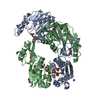
Deposited unit
A: EPHRIN TYPE-B RECEPTOR 2
B: EPHRIN TYPE-B RECEPTOR 2
C: EPHRIN TYPE-B RECEPTOR 2
D: EPHRIN TYPE-B RECEPTOR 2
E: EPHRIN-B2
F: EPHRIN-B2
G: EPHRIN-B2
H: EPHRIN-B2


Theoretical massNumber of molelcules
Total (without water)146,2908
Polymers146,2908
Non-polymers00
Water00
1
A: EPHRIN TYPE-B RECEPTOR 2
D: EPHRIN TYPE-B RECEPTOR 2
E: EPHRIN-B2
H: EPHRIN-B2


Theoretical massNumber of molelcules
Total (without water)73,1454
Polymers73,1454
Non-polymers00
Water0
TypeNameSymmetry operationNumber
identity operation1_555x,y,z1
2
B: EPHRIN TYPE-B RECEPTOR 2
C: EPHRIN TYPE-B RECEPTOR 2
F: EPHRIN-B2
G: EPHRIN-B2


Theoretical massNumber of molelcules
Total (without water)73,1454
Polymers73,1454
Non-polymers00
Water0
TypeNameSymmetry operationNumber
identity operation1_555x,y,z1
Buried area6950 Å2
ΔGint-36 kcal/mol
Surface area28360 Å2
MethodPISA
Unit cell
Length a, b, c (Å)78, 78, 78
Angle α, β, γ (deg.)69, 75, 69
Int Tables number1
Space group name H-MP1
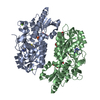
Detailschains B and F form a heterodimer chains C and D form a heterodimer Chains B, C, F, and G form a proposed heterotetramer

-
Components

#1: Protein
EPHRIN TYPE-B RECEPTOR 2


Mass: 20856.512 Da / Num. of mol.: 4 / Fragment: ligand-binding domain
Source method: isolated from a genetically manipulated source
Source: (gene. exp.) Mus musculus (house mouse) / Production host: Escherichia coli (E. coli) / References: UniProt: P54763, EC: 2.7.1.112
#2: Protein
EPHRIN-B2


Mass: 15715.984 Da / Num. of mol.: 4 / Fragment: extracellular domain
Source method: isolated from a genetically manipulated source
Source: (gene. exp.) Mus musculus (house mouse) / Production host: Escherichia coli (E. coli) / References: UniProt: P52800
Has protein modificationY

-
Experimental details

-
Experiment

ExperimentMethod: X-RAY DIFFRACTION / Number of used crystals: 1

-
Sample preparation

CrystalDensity Matthews: 2.79 Å3/Da / Density % sol: 55.98 %
Crystal growTemperature: 293 K / Method: vapor diffusion, hanging drop / pH: 7.5
Details: 1.6 M Na/K Phosphate, 100 mM HEPES, pH 7.5, VAPOR DIFFUSION, HANGING DROP, temperature 293K
Crystal grow
*PLUS
Temperature: 20 ℃ / pH: 7.2
Components of the solutions
*PLUS
IDConc.Common nameCrystal-IDSol-IDChemical formulaDetails (eV)
120 mg/mlprotein1drop
210 mM1dropKCl
32 mM1dropMgCl2
410 mMHEPES1droppH7.2
51.6 Msodium potassium phospahte1reservoir
6100 mMHEPES-NaOH1reservoirpH7.5

-
Data collection

DiffractionMean temperature: 123 K
Diffraction sourceSource: SYNCHROTRON / Site: CHESS / Beamline: F2 / Wavelength: 0.98 Å
DetectorType: ADSC QUANTUM 4 / Detector: CCD / Date: Sep 1, 2000
RadiationMonochromator: Si / Protocol: MAD / Monochromatic (M) / Laue (L): M / Scattering type: x-ray
Radiation wavelengthWavelength: 0.98 Å / Relative weight: 1
ReflectionResolution: 2.7→25 Å / Num. all: 43549 / Num. obs: 42156 / % possible obs: 96.8 % / Observed criterion σ(F): 1 / Observed criterion σ(I): 1 / Rmerge(I) obs: 0.073
Reflection shellResolution: 2.7→2.82 Å / Rmerge(I) obs: 0.37 / % possible all: 93.5

-
Processing

Software
NameClassification
MAR345data collection
SCALEPACKdata scaling
MLPHAREphasing
CNSrefinement
RefinementMethod to determine structure: MAD / Resolution: 2.7→25 Å / σ(F): 1 / Stereochemistry target values: Engh & Huber
RfactorNum. reflectionSelection details
Rfree0.2956 2581 random
Rwork0.2427 --
all-43465 -
obs-42787 -
Refinement stepCycle: LAST / Resolution: 2.7→25 Å
ProteinNucleic acidLigandSolventTotal
Num. atoms10260 0 0 0 10260
Refine LS restraints
Refine-IDTypeDev ideal
X-RAY DIFFRACTIONx_bond_d0.0082
X-RAY DIFFRACTIONx_angle_deg1.6374
Refinement
*PLUS
Rfactor obs: 0.243 / Rfactor Rfree: 0.295
Solvent computation
*PLUS
Displacement parameters
*PLUS

+
About Yorodumi

-
News

-
Feb 9, 2022. New format data for meta-information of EMDB entries

New format data for meta-information of EMDB entries

  • Version 3 of the EMDB header file is now the official format.
  • The previous official version 1.9 will be removed from the archive.

Related info.:EMDB header

External links:wwPDB to switch to version 3 of the EMDB data model

-
Aug 12, 2020. Covid-19 info

Covid-19 info

URL: https://pdbj.org/emnavi/covid19.php

New page: Covid-19 featured information page in EM Navigator.

Related info.:Covid-19 info / Mar 5, 2020. Novel coronavirus structure data

+
Mar 5, 2020. Novel coronavirus structure data

Novel coronavirus structure data

Related info.:Yorodumi Speices / Aug 12, 2020. Covid-19 info

External links:COVID-19 featured content - PDBj / Molecule of the Month (242):Coronavirus Proteases

+
Jan 31, 2019. EMDB accession codes are about to change! (news from PDBe EMDB page)

EMDB accession codes are about to change! (news from PDBe EMDB page)

  • The allocation of 4 digits for EMDB accession codes will soon come to an end. Whilst these codes will remain in use, new EMDB accession codes will include an additional digit and will expand incrementally as the available range of codes is exhausted. The current 4-digit format prefixed with “EMD-” (i.e. EMD-XXXX) will advance to a 5-digit format (i.e. EMD-XXXXX), and so on. It is currently estimated that the 4-digit codes will be depleted around Spring 2019, at which point the 5-digit format will come into force.
  • The EM Navigator/Yorodumi systems omit the EMD- prefix.

Related info.:Q: What is EMD? / ID/Accession-code notation in Yorodumi/EM Navigator

External links:EMDB Accession Codes are Changing Soon! / Contact to PDBj

+
Jul 12, 2017. Major update of PDB

Major update of PDB

  • wwPDB released updated PDB data conforming to the new PDBx/mmCIF dictionary.
  • This is a major update changing the version number from 4 to 5, and with Remediation, in which all the entries are updated.
  • In this update, many items about electron microscopy experimental information are reorganized (e.g. em_software).
  • Now, EM Navigator and Yorodumi are based on the updated data.

External links:wwPDB Remediation / Enriched Model Files Conforming to OneDep Data Standards Now Available in the PDB FTP Archive

-
Yorodumi

Thousand views of thousand structures

  • Yorodumi is a browser for structure data from EMDB, PDB, SASBDB, etc.
  • This page is also the successor to EM Navigator detail page, and also detail information page/front-end page for Omokage search.
  • The word "yorodu" (or yorozu) is an old Japanese word meaning "ten thousand". "mi" (miru) is to see.

Related info.:EMDB / PDB / SASBDB / Comparison of 3 databanks / Yorodumi Search / Aug 31, 2016. New EM Navigator & Yorodumi / Yorodumi Papers / Jmol/JSmol / Function and homology information / Changes in new EM Navigator and Yorodumi

Read more