- PDB-2gw5: Crystal Structure of LIR-2 (ILT4) at 1.8 : differences from LIR-1... -
+
Open data
ID or keywords:
Loading...
-
Basic information
Entry
Database: PDB / ID: 2gw5
Title
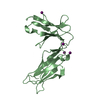
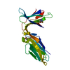
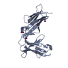
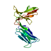
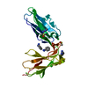
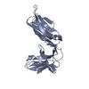
Crystal Structure of LIR-2 (ILT4) at 1.8 : differences from LIR-1 (ILT2) in regions implicated in the binding of the Cytomegalovirus class I MHC homolog UL18
Components
Leukocyte immunoglobulin-like receptor subfamily B member 2 precursor
Keywords
IMMUNE SYSTEM / Ig like domains
Function / homology
Function and homology information
negative regulation of antigen processing and presentation / negative regulation of T cell costimulation / positive regulation of tolerance induction / MHC class Ib protein complex binding / inhibitory MHC class I receptor activity / Fc receptor mediated inhibitory signaling pathway / negative regulation of postsynaptic density organization / immune response-inhibiting cell surface receptor signaling pathway / positive regulation of long-term synaptic depression / MHC class Ib protein binding ...negative regulation of antigen processing and presentation / negative regulation of T cell costimulation / positive regulation of tolerance induction / MHC class Ib protein complex binding / inhibitory MHC class I receptor activity / Fc receptor mediated inhibitory signaling pathway / negative regulation of postsynaptic density organization / immune response-inhibiting cell surface receptor signaling pathway / positive regulation of long-term synaptic depression / MHC class Ib protein binding / immune response-regulating signaling pathway / positive regulation of T cell tolerance induction / interleukin-10-mediated signaling pathway / protein phosphatase 1 binding / regulation of dendritic cell differentiation / regulation of long-term synaptic potentiation / negative regulation of protein metabolic process / positive regulation of regulatory T cell differentiation / heterotypic cell-cell adhesion / negative regulation of calcium ion transport / tertiary granule membrane / ficolin-1-rich granule membrane / MHC class I protein binding / cellular defense response / negative regulation of T cell proliferation / positive regulation of T cell proliferation / cell adhesion molecule binding / positive regulation of interleukin-6 production / Immunoregulatory interactions between a Lymphoid and a non-Lymphoid cell / cell-cell signaling / amyloid-beta binding / cellular response to lipopolysaccharide / adaptive immune response / learning or memory / cell surface receptor signaling pathway / immune response / Neutrophil degranulation / protein-containing complex binding / cell surface / signal transduction / protein homodimerization activity / : / membrane / plasma membrane / cytoplasm Similarity search - Function
Resolution: 1.8→34.08 Å / Cor.coef. Fo:Fc: 0.931 / Cor.coef. Fo:Fc free: 0.915 / SU B: 3.2 / SU ML: 0.102 / Cross valid method: THROUGHOUT / ESU R: 0.152 / ESU R Free: 0.145 / Stereochemistry target values: MAXIMUM LIKELIHOOD / Details: HYDROGENS HAVE BEEN ADDED IN THE RIDING POSITIONS
Rfactor
Num. reflection
% reflection
Selection details
Rfree
0.27864
991
4.9 %
RANDOM
Rwork
0.24041
-
-
-
obs
0.2423
19239
99.38 %
-
Solvent computation
Ion probe radii: 0.8 Å / Shrinkage radii: 0.8 Å / VDW probe radii: 1.4 Å / Solvent model: MASK
Displacement parameters
Biso mean: 28.611 Å2
Baniso -1
Baniso -2
Baniso -3
1-
0 Å2
0 Å2
0 Å2
2-
-
0 Å2
0 Å2
3-
-
-
-0.01 Å2
Refinement step
Cycle: LAST / Resolution: 1.8→34.08 Å
Protein
Nucleic acid
Ligand
Solvent
Total
Num. atoms
1398
0
4
145
1547
Refine LS restraints
Refine-ID
Type
Dev ideal
Dev ideal target
Number
X-RAY DIFFRACTION
r_bond_refined_d
0.026
0.022
1453
X-RAY DIFFRACTION
r_bond_other_d
X-RAY DIFFRACTION
r_angle_refined_deg
2.162
1.968
1989
X-RAY DIFFRACTION
r_angle_other_deg
X-RAY DIFFRACTION
r_dihedral_angle_1_deg
7.262
5
184
X-RAY DIFFRACTION
r_dihedral_angle_2_deg
33.96
22.364
55
X-RAY DIFFRACTION
r_dihedral_angle_3_deg
16.954
15
209
X-RAY DIFFRACTION
r_dihedral_angle_4_deg
20.317
15
10
X-RAY DIFFRACTION
r_chiral_restr
0.161
0.2
218
X-RAY DIFFRACTION
r_gen_planes_refined
0.01
0.02
1112
X-RAY DIFFRACTION
r_gen_planes_other
X-RAY DIFFRACTION
r_nbd_refined
0.314
0.2
656
X-RAY DIFFRACTION
r_nbd_other
X-RAY DIFFRACTION
r_nbtor_refined
0.319
0.2
945
X-RAY DIFFRACTION
r_nbtor_other
X-RAY DIFFRACTION
r_xyhbond_nbd_refined
0.206
0.2
128
X-RAY DIFFRACTION
r_xyhbond_nbd_other
X-RAY DIFFRACTION
r_metal_ion_refined
X-RAY DIFFRACTION
r_metal_ion_other
X-RAY DIFFRACTION
r_symmetry_vdw_refined
0.3
0.2
26
X-RAY DIFFRACTION
r_symmetry_vdw_other
X-RAY DIFFRACTION
r_symmetry_hbond_refined
0.368
0.2
9
X-RAY DIFFRACTION
r_symmetry_hbond_other
X-RAY DIFFRACTION
r_symmetry_metal_ion_refined
X-RAY DIFFRACTION
r_symmetry_metal_ion_other
X-RAY DIFFRACTION
r_mcbond_it
1.623
1.5
962
X-RAY DIFFRACTION
r_mcbond_other
X-RAY DIFFRACTION
r_mcangle_it
2.451
2
1491
X-RAY DIFFRACTION
r_scbond_it
3.865
3
595
X-RAY DIFFRACTION
r_scangle_it
5.362
4.5
496
X-RAY DIFFRACTION
r_rigid_bond_restr
X-RAY DIFFRACTION
r_sphericity_free
X-RAY DIFFRACTION
r_sphericity_bonded
LS refinement shell
Resolution: 1.8→1.847 Å / Total num. of bins used: 20
Rfactor
Num. reflection
% reflection
Rfree
0.373
89
-
Rwork
0.294
1398
-
obs
-
-
100 %
+
About Yorodumi
-
News
-
Feb 9, 2022. New format data for meta-information of EMDB entries
New format data for meta-information of EMDB entries
Version 3 of the EMDB header file is now the official format.
The previous official version 1.9 will be removed from the archive.
In the structure databanks used in Yorodumi, some data are registered as the other names, "COVID-19 virus" and "2019-nCoV". Here are the details of the virus and the list of structure data.
Jan 31, 2019. EMDB accession codes are about to change! (news from PDBe EMDB page)
EMDB accession codes are about to change! (news from PDBe EMDB page)
The allocation of 4 digits for EMDB accession codes will soon come to an end. Whilst these codes will remain in use, new EMDB accession codes will include an additional digit and will expand incrementally as the available range of codes is exhausted. The current 4-digit format prefixed with “EMD-” (i.e. EMD-XXXX) will advance to a 5-digit format (i.e. EMD-XXXXX), and so on. It is currently estimated that the 4-digit codes will be depleted around Spring 2019, at which point the 5-digit format will come into force.
The EM Navigator/Yorodumi systems omit the EMD- prefix.
Related info.:Q: What is EMD? / ID/Accession-code notation in Yorodumi/EM Navigator
Yorodumi is a browser for structure data from EMDB, PDB, SASBDB, etc.
This page is also the successor to EM Navigator detail page, and also detail information page/front-end page for Omokage search.
The word "yorodu" (or yorozu) is an old Japanese word meaning "ten thousand". "mi" (miru) is to see.
Related info.:EMDB / PDB / SASBDB / Comparison of 3 databanks / Yorodumi Search / Aug 31, 2016. New EM Navigator & Yorodumi / Yorodumi Papers / Jmol/JSmol / Function and homology information / Changes in new EM Navigator and Yorodumi
 Movie
Movie Controller

 Yorodumi
 Open data
 Basic information
 Components
 Keywords
 Function and homology information
 Homo sapiens (human)
 X-RAY DIFFRACTION /
  Authors
 Citation
 Structure visualization
 Downloads & links
 Other downloads
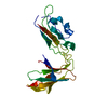


 PDBj
PDBj


 Assembly
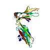


 Mass: 60.095 Da / Num. of mol.: 1 / Source method: obtained synthetically / Formula: C3H8O
 Mass: 18.015 Da / Num. of mol.: 145 / Source method: isolated from a natural source / Formula: H2O
 Sample preparation
 / Beamline: BL9-2 / Wavelength: 0.999 Å
 Processing