 Movie
Movie Controller


+ Open data

- Basic information
| Entry | Database: PDB / ID: 1g0x | ||||||
|---|---|---|---|---|---|---|---|
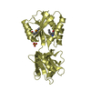
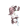
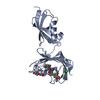
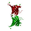
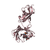
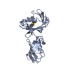
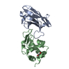
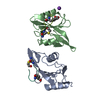
| Title | CRYSTAL STRUCTURE OF THE LIGAND BINDING DOMAIN OF LIR-1 (ILT2) | ||||||
 Components Components | LEUCOCYTE IMMUNOGLOBULIN-LIKE RECEPTOR-1 | ||||||
 Keywords Keywords | IMMUNE SYSTEM / Immunoglobulin fold / 3-10 helix | ||||||
| Function / homology |  Function and homology information Function and homology informationHLA-A specific inhibitory MHC class I receptor activity / : / negative regulation of serotonin secretion / MHC class Ib protein complex binding / HLA-B specific inhibitory MHC class I receptor activity / inhibitory MHC class I receptor activity / negative regulation of dendritic cell differentiation / immune response-inhibiting cell surface receptor signaling pathway / Fc receptor mediated inhibitory signaling pathway / MHC class Ib receptor activity ...HLA-A specific inhibitory MHC class I receptor activity / : / negative regulation of serotonin secretion / MHC class Ib protein complex binding / HLA-B specific inhibitory MHC class I receptor activity / inhibitory MHC class I receptor activity / negative regulation of dendritic cell differentiation / immune response-inhibiting cell surface receptor signaling pathway / Fc receptor mediated inhibitory signaling pathway / MHC class Ib receptor activity / MHC class Ib protein binding / negative regulation of T cell mediated cytotoxicity / immune response-regulating signaling pathway / negative regulation of CD8-positive, alpha-beta T cell activation / negative regulation of transforming growth factor beta production / MHC class I receptor activity / negative regulation of cytokine production involved in immune response / dendritic cell differentiation / interleukin-10-mediated signaling pathway / negative regulation of osteoclast development / negative regulation of T cell activation via T cell receptor contact with antigen bound to MHC molecule on antigen presenting cell / protein phosphatase 1 binding / negative regulation of interleukin-12 production / negative regulation of alpha-beta T cell activation / negative regulation of dendritic cell apoptotic process / negative regulation of endocytosis / negative regulation of interferon-beta production / negative regulation of mononuclear cell proliferation / negative regulation of natural killer cell mediated cytotoxicity / T cell proliferation involved in immune response / positive regulation of macrophage cytokine production / negative regulation of interleukin-10 production / negative regulation of type II interferon production / negative regulation of calcium ion transport / negative regulation of cell cycle / negative regulation of tumor necrosis factor production / MHC class I protein binding / negative regulation of T cell proliferation / positive regulation of defense response to virus by host / SH2 domain binding / receptor internalization / positive regulation of type II interferon production / Immunoregulatory interactions between a Lymphoid and a non-Lymphoid cell / response to virus / cellular response to lipopolysaccharide / defense response to virus / adaptive immune response / positive regulation of apoptotic process / external side of plasma membrane / positive regulation of gene expression / signal transduction / protein homodimerization activity / positive regulation of transcription by RNA polymerase II / extracellular region / plasma membrane / cytoplasm Similarity search - Function | ||||||
| Biological species |  Homo sapiens (human) Homo sapiens (human) | ||||||
| Method |  X-RAY DIFFRACTION / SYNCHROTRON / Resolution: 2.1 Å X-RAY DIFFRACTION / SYNCHROTRON / Resolution: 2.1 Å | ||||||
 Authors Authors | Chapman, T.L. / Heikema, A.P. / West Jr., A.P. / Bjorkman, P.J. | ||||||
 Citation Citation | Journal: Immunity / Year: 2000 Title: Crystal structure and ligand binding properties of the D1D2 region of the inhibitory receptor LIR-1 (ILT2). Authors: Chapman, T.L. / Heikema, A.P. / West Jr., A.P. / Bjorkman, P.J. | ||||||
| History |
|
- Structure visualization
| Structure viewer | Molecule: MolmilJmol/JSmol |
|---|
- Downloads & links
-Download
| PDBx/mmCIF format | 1g0x.cif.gz | 55.6 KB | Display | PDBx/mmCIF format |
|---|---|---|---|---|
| PDB format | pdb1g0x.ent.gz | 40 KB | Display | PDB format |
| PDBx/mmJSON format | 1g0x.json.gz | Tree view | PDBx/mmJSON format | |
| Others |  Other downloads Other downloads |
-Validation report
| Arichive directory | https://data.pdbj.org/pub/pdb/validation_reports/g0/1g0xftp://data.pdbj.org/pub/pdb/validation_reports/g0/1g0x | HTTPS FTP |
|---|
-Related structure data
| Similar structure data |
|---|

-Links
 PDBj
PDBj


- Assembly
| Deposited unit | 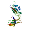
| ||||||||
|---|---|---|---|---|---|---|---|---|---|
| 1 |
| ||||||||
| Unit cell |
|
-Components
| #1: Protein | Mass: 21998.645 Da / Num. of mol.: 1 / Fragment: D1D2 LIGAND BINDING DOMAIN Source method: isolated from a genetically manipulated source Source: (gene. exp.) Homo sapiens (human) / Plasmid: PET23 / Production host:  |
|---|---|
| #2: Water | ChemComp-HOH /  Mass: 18.015 Da / Num. of mol.: 245 / Source method: isolated from a natural source / Formula: H2O Mass: 18.015 Da / Num. of mol.: 245 / Source method: isolated from a natural source / Formula: H2O |
| Has protein modification | Y |
-Experimental details
-Experiment
| Experiment | Method: X-RAY DIFFRACTION / Number of used crystals: 1 |
|---|
- Sample preparation
| Crystal | Density Matthews: 3.43 Å3/Da / Density % sol: 64.16 % | ||||||||||||||||||||||||
|---|---|---|---|---|---|---|---|---|---|---|---|---|---|---|---|---|---|---|---|---|---|---|---|---|---|
| Crystal grow | Temperature: 298 K / Method: vapor diffusion, hanging drop / pH: 8.5 Details: potassium sodium tartarate, tris, pH 8.5, VAPOR DIFFUSION, HANGING DROP, temperature 298K | ||||||||||||||||||||||||
| Crystal grow | *PLUS Temperature: 22 ℃ | ||||||||||||||||||||||||
| Components of the solutions | *PLUS
|
-Data collection
| Diffraction | Mean temperature: 100 K |
|---|---|
| Diffraction source | Source: SYNCHROTRON / Site: SSRL  / Beamline: BL9-2 / Wavelength: 1 / Beamline: BL9-2 / Wavelength: 1 |
| Detector | Type: ADSC QUANTUM 4 / Detector: CCD / Date: Dec 1, 1999 |
| Radiation | Protocol: SINGLE WAVELENGTH / Monochromatic (M) / Laue (L): M / Scattering type: x-ray |
| Radiation wavelength | Wavelength: 1 Å / Relative weight: 1 |
| Reflection | Resolution: 2.1→20 Å / Num. obs: 21555 / % possible obs: 99.5 % / Observed criterion σ(I): -3 / Redundancy: 3.6 % / Biso Wilson estimate: 16.2 Å2 / Rmerge(I) obs: 0.06 / Net I/σ(I): 18.8 |
| Reflection shell | Resolution: 2.1→2.17 Å / Redundancy: 3.5 % / Rmerge(I) obs: 0.297 / Num. unique all: 2112 / % possible all: 99.5 |
| Reflection | *PLUS Lowest resolution: 30 Å / Rmerge(I) obs: 0.06 |
| Reflection shell | *PLUS % possible obs: 99.5 % / Num. unique obs: 2112 / Mean I/σ(I) obs: 3.6 |
- Processing
| Software |
| ||||||||||||||||||||||||||||||||||||||||
|---|---|---|---|---|---|---|---|---|---|---|---|---|---|---|---|---|---|---|---|---|---|---|---|---|---|---|---|---|---|---|---|---|---|---|---|---|---|---|---|---|---|
| Refinement | Resolution: 2.1→19.77 Å / Rfactor Rfree error: 0.008 / Data cutoff high absF: 1902676.83 / Data cutoff low absF: 0 / Isotropic thermal model: RESTRAINED / Cross valid method: THROUGHOUT / σ(F): 0
| ||||||||||||||||||||||||||||||||||||||||
| Solvent computation | Solvent model: FLAT MODEL / Bsol: 49.83 Å2 / ksol: 0.361 e/Å3 | ||||||||||||||||||||||||||||||||||||||||
| Displacement parameters | Biso mean: 29.6 Å2
| ||||||||||||||||||||||||||||||||||||||||
| Refine analyze |
| ||||||||||||||||||||||||||||||||||||||||
| Refinement step | Cycle: LAST / Resolution: 2.1→19.77 Å
| ||||||||||||||||||||||||||||||||||||||||
| Refine LS restraints |
| ||||||||||||||||||||||||||||||||||||||||
| LS refinement shell | Resolution: 2.1→2.17 Å / Rfactor Rfree error: 0.029 / Total num. of bins used: 10
| ||||||||||||||||||||||||||||||||||||||||
| Xplor file |
| ||||||||||||||||||||||||||||||||||||||||
| Software | *PLUS Name: CNS / Version: 1 / Classification: refinement | ||||||||||||||||||||||||||||||||||||||||
| Refinement | *PLUS σ(F): 0 / % reflection Rfree: 4.9 % | ||||||||||||||||||||||||||||||||||||||||
| Solvent computation | *PLUS | ||||||||||||||||||||||||||||||||||||||||
| Displacement parameters | *PLUS Biso mean: 29.6 Å2 | ||||||||||||||||||||||||||||||||||||||||
| Refine LS restraints | *PLUS
| ||||||||||||||||||||||||||||||||||||||||
| LS refinement shell | *PLUS Rfactor Rfree: 0.287 / % reflection Rfree: 5.5 % / Rfactor Rwork: 0.226 |
