 Movie
Movie Controller

[English] 日本語
 Yorodumi
- PDB-4u3x: Structure of a human VH antibody domain binding to the cleft of h... -
+ Open data

- Basic information
| Entry | Database: PDB / ID: 4u3x | ||||||
|---|---|---|---|---|---|---|---|
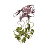
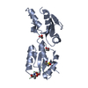
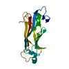
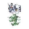
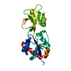
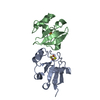
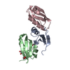
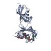
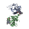
| Title | Structure of a human VH antibody domain binding to the cleft of hen egg lysozyme | ||||||
 Components Components |
| ||||||
 Keywords Keywords | IMMUNE SYSTEM / Human VH domain antibody / Phage display / Antibody-antigen complex structure / HYDROLASE-IMMUNE SYSTEM complex | ||||||
| Function / homology |  Function and homology information Function and homology informationLactose synthesis / Antimicrobial peptides / Neutrophil degranulation / beta-N-acetylglucosaminidase activity / cell wall macromolecule catabolic process / lysozyme / lysozyme activity / killing of cells of another organism / defense response to Gram-negative bacterium / defense response to bacterium ...Lactose synthesis / Antimicrobial peptides / Neutrophil degranulation / beta-N-acetylglucosaminidase activity / cell wall macromolecule catabolic process / lysozyme / lysozyme activity / killing of cells of another organism / defense response to Gram-negative bacterium / defense response to bacterium / defense response to Gram-positive bacterium / endoplasmic reticulum / : / identical protein binding / cytoplasm Similarity search - Function | ||||||
| Biological species |  Homo sapiens (human) Homo sapiens (human) | ||||||
| Method |  X-RAY DIFFRACTION / SYNCHROTRON / Resolution: 2.26 Å X-RAY DIFFRACTION / SYNCHROTRON / Resolution: 2.26 Å | ||||||
 Authors Authors | Rouet, R. / Langley, D.B. / Christ, D. | ||||||
 Citation Citation | Journal: J.Biol.Chem. / Year: 2015 Title: Fully Human VH Single Domains That Rival the Stability and Cleft Recognition of Camelid Antibodies Authors: Rouet, R. / Dudgeon, K. / Christie, M. / Langley, D. / Christ, D. | ||||||
| History |
|
- Structure visualization
| Structure viewer | Molecule: MolmilJmol/JSmol |
|---|
- Downloads & links
-Download
| PDBx/mmCIF format | 4u3x.cif.gz | 190.2 KB | Display | PDBx/mmCIF format |
|---|---|---|---|---|
| PDB format | pdb4u3x.ent.gz | 152.4 KB | Display | PDB format |
| PDBx/mmJSON format | 4u3x.json.gz | Tree view | PDBx/mmJSON format | |
| Others |  Other downloads Other downloads |
-Validation report
| Arichive directory | https://data.pdbj.org/pub/pdb/validation_reports/u3/4u3xftp://data.pdbj.org/pub/pdb/validation_reports/u3/4u3x | HTTPS FTP |
|---|
-Related structure data
| Related structure data | 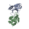 4pgjC 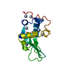 1lyzS  1ohqS C: citing same article ( S: Starting model for refinement |
|---|---|
| Similar structure data |

-Links
 PDBj
PDBj


- Assembly
| Deposited unit | 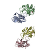
| ||||||||
|---|---|---|---|---|---|---|---|---|---|
| 1 | 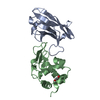
| ||||||||
| 2 | 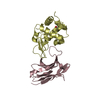
| ||||||||
| Unit cell |
|
-Components
| #1: Antibody | Mass: 13173.522 Da / Num. of mol.: 2 Source method: isolated from a genetically manipulated source Source: (gene. exp.) Homo sapiens (human) / Production host:  #2: Protein | Mass: 14331.160 Da / Num. of mol.: 2 / Source method: isolated from a natural source / Source: (natural) #3: Chemical |   Mass: 62.068 Da / Num. of mol.: 2 / Source method: isolated from a natural source / Formula: C2H6O2 Mass: 62.068 Da / Num. of mol.: 2 / Source method: isolated from a natural source / Formula: C2H6O2#4: Water | ChemComp-HOH / |  Mass: 18.015 Da / Num. of mol.: 101 / Source method: isolated from a natural source / Formula: H2O Mass: 18.015 Da / Num. of mol.: 101 / Source method: isolated from a natural source / Formula: H2OHas protein modification | Y | |
|---|
-Experimental details
-Experiment
| Experiment | Method: X-RAY DIFFRACTION |
|---|
- Sample preparation
| Crystal | Density Matthews: 2.32 Å3/Da / Density % sol: 46.94 % |
|---|---|
| Crystal grow | Temperature: 298 K / Method: vapor diffusion, hanging drop / pH: 5.5 / Details: 300 mM sodium citrate, 16% PEG 3350 / PH range: 5.5 |
-Data collection
| Diffraction | Mean temperature: 100 K |
|---|---|
| Diffraction source | Source: SYNCHROTRON / Site: Australian Synchrotron  / Beamline: MX2 / Wavelength: 0.9537 Å / Beamline: MX2 / Wavelength: 0.9537 Å |
| Detector | Type: ADSC QUANTUM 315r / Detector: CCD / Date: Apr 12, 2014 |
| Radiation | Protocol: SINGLE WAVELENGTH / Monochromatic (M) / Laue (L): M / Scattering type: x-ray |
| Radiation wavelength | Wavelength: 0.9537 Å / Relative weight: 1 |
| Reflection | Resolution: 2.26→97.14 Å / Num. obs: 24456 / % possible obs: 98.6 % / Redundancy: 1.5 % / Net I/σ(I): 2.4 |
- Processing
| Software | Name: REFMAC / Version: 5.8.0071 / Classification: refinement | ||||||||||||||||||||||||||||||||||||||||||||||||||||||||||||||||||||||||||||||||||||||||||||||||||||||||||||||||||||||||||||||||||||||||||||||||||||||||||||||||||||||||||||||||||||||
|---|---|---|---|---|---|---|---|---|---|---|---|---|---|---|---|---|---|---|---|---|---|---|---|---|---|---|---|---|---|---|---|---|---|---|---|---|---|---|---|---|---|---|---|---|---|---|---|---|---|---|---|---|---|---|---|---|---|---|---|---|---|---|---|---|---|---|---|---|---|---|---|---|---|---|---|---|---|---|---|---|---|---|---|---|---|---|---|---|---|---|---|---|---|---|---|---|---|---|---|---|---|---|---|---|---|---|---|---|---|---|---|---|---|---|---|---|---|---|---|---|---|---|---|---|---|---|---|---|---|---|---|---|---|---|---|---|---|---|---|---|---|---|---|---|---|---|---|---|---|---|---|---|---|---|---|---|---|---|---|---|---|---|---|---|---|---|---|---|---|---|---|---|---|---|---|---|---|---|---|---|---|---|---|
| Refinement | Starting model: 1LYZ, 1OHQ Resolution: 2.26→97.14 Å / Cor.coef. Fo:Fc: 0.942 / Cor.coef. Fo:Fc free: 0.892 / SU B: 15.494 / SU ML: 0.18 / Cross valid method: THROUGHOUT / ESU R: 0.298 / ESU R Free: 0.232 / Stereochemistry target values: MAXIMUM LIKELIHOOD
| ||||||||||||||||||||||||||||||||||||||||||||||||||||||||||||||||||||||||||||||||||||||||||||||||||||||||||||||||||||||||||||||||||||||||||||||||||||||||||||||||||||||||||||||||||||||
| Solvent computation | Ion probe radii: 0.8 Å / Shrinkage radii: 0.8 Å / VDW probe radii: 1.2 Å / Solvent model: MASK | ||||||||||||||||||||||||||||||||||||||||||||||||||||||||||||||||||||||||||||||||||||||||||||||||||||||||||||||||||||||||||||||||||||||||||||||||||||||||||||||||||||||||||||||||||||||
| Displacement parameters | Biso mean: 14.923 Å2
| ||||||||||||||||||||||||||||||||||||||||||||||||||||||||||||||||||||||||||||||||||||||||||||||||||||||||||||||||||||||||||||||||||||||||||||||||||||||||||||||||||||||||||||||||||||||
| Refinement step | Cycle: 1 / Resolution: 2.26→97.14 Å
| ||||||||||||||||||||||||||||||||||||||||||||||||||||||||||||||||||||||||||||||||||||||||||||||||||||||||||||||||||||||||||||||||||||||||||||||||||||||||||||||||||||||||||||||||||||||
| Refine LS restraints |
|