 Movie
Movie Controller


+ Open data

- Basic information
| Entry | Database: PDB / ID: 3h8n | ||||||
|---|---|---|---|---|---|---|---|
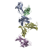
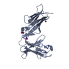
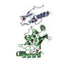
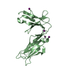
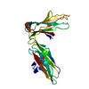
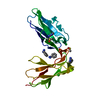
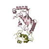
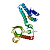
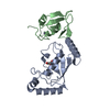
| Title | Crystal Structure Analysis of KIR2DS4 | ||||||
 Components Components | Killer cell immunoglobulin-like receptor 2DS4 | ||||||
 Keywords Keywords | IMMUNE SYSTEM / ligand-binding domains / Cell membrane / Disulfide bond / Glycoprotein / Immunoglobulin domain / Membrane / Polymorphism / Receptor / Transmembrane | ||||||
| Function / homology |  Function and homology information Function and homology informationMHC class Ib protein binding / immune response-regulating signaling pathway / DAP12 interactions / plasma membrane Similarity search - Function | ||||||
| Biological species |  Homo sapiens (human) Homo sapiens (human) | ||||||
| Method |  X-RAY DIFFRACTION / SYNCHROTRON / MOLECULAR REPLACEMENT / Resolution: 2.5 Å X-RAY DIFFRACTION / SYNCHROTRON / MOLECULAR REPLACEMENT / Resolution: 2.5 Å | ||||||
 Authors Authors | Graef, T. / Bushnell, D.A. / Parham, P. | ||||||
 Citation Citation | Journal: J.Exp.Med. / Year: 2009 Title: KIR2DS4 is a product of gene conversion with KIR3DL2 that introduced specificity for HLA-A*11 while diminishing avidity for HLA-C. Authors: Graef, T. / Moesta, A.K. / Norman, P.J. / Abi-Rached, L. / Vago, L. / Older Aguilar, A.M. / Gleimer, M. / Hammond, J.A. / Guethlein, L.A. / Bushnell, D.A. / Robinson, P.J. / Parham, P. | ||||||
| History |
|
- Structure visualization
| Structure viewer | Molecule: MolmilJmol/JSmol |
|---|
- Downloads & links
-Download
| PDBx/mmCIF format | 3h8n.cif.gz | 52.2 KB | Display | PDBx/mmCIF format |
|---|---|---|---|---|
| PDB format | pdb3h8n.ent.gz | 36.9 KB | Display | PDB format |
| PDBx/mmJSON format | 3h8n.json.gz | Tree view | PDBx/mmJSON format | |
| Others |  Other downloads Other downloads |
-Validation report
| Arichive directory | https://data.pdbj.org/pub/pdb/validation_reports/h8/3h8nftp://data.pdbj.org/pub/pdb/validation_reports/h8/3h8n | HTTPS FTP |
|---|
-Related structure data
| Related structure data | 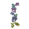 1im9S S: Starting model for refinement |
|---|---|
| Similar structure data |


-Links
 PDBj
PDBj


- Assembly
| Deposited unit | 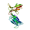
| ||||||||
|---|---|---|---|---|---|---|---|---|---|
| 1 |
| ||||||||
| Unit cell |
|
-Components
| #1: Protein | Mass: 21534.236 Da / Num. of mol.: 1 Source method: isolated from a genetically manipulated source Source: (gene. exp.) Homo sapiens (human) / Gene: CD158I, KIR2DS4, KIR2DS4*0010101, KKA3, NKAT8 / Plasmid: pET-28c / Production host:  |
|---|---|
| #2: Water | ChemComp-HOH /  Mass: 18.015 Da / Num. of mol.: 53 / Source method: isolated from a natural source / Formula: H2O Mass: 18.015 Da / Num. of mol.: 53 / Source method: isolated from a natural source / Formula: H2O |
| Has protein modification | Y |
-Experimental details
-Experiment
| Experiment | Method: X-RAY DIFFRACTION / Number of used crystals: 1 |
|---|
- Sample preparation
| Crystal | Density Matthews: 2.46 Å3/Da / Density % sol: 50.1 % |
|---|---|
| Crystal grow | Temperature: 283 K / Method: vapor diffusion, hanging drop / pH: 8.5 Details: 0.22 M Mg-Formate, 100 mM BIS-TRIS, pH 5.5, VAPOR DIFFUSION, HANGING DROP, temperature 283K |
-Data collection
| Diffraction | Mean temperature: 100 K |
|---|---|
| Diffraction source | Source: SYNCHROTRON / Site: SSRL  / Beamline: BL11-1 / Wavelength: 0.979 Å / Beamline: BL11-1 / Wavelength: 0.979 Å |
| Detector | Type: MARMOSAIC 325 mm CCD / Detector: CCD / Date: Jan 7, 2008 |
| Radiation | Protocol: SINGLE WAVELENGTH / Monochromatic (M) / Laue (L): M / Scattering type: x-ray |
| Radiation wavelength | Wavelength: 0.979 Å / Relative weight: 1 |
| Reflection | Resolution: 2.5→40.45 Å / Num. all: 7814 / Num. obs: 7795 / % possible obs: 99.8 % / Observed criterion σ(I): -3 / Redundancy: 13.7 % / Biso Wilson estimate: 44.26 Å2 / Rsym value: 0.05 / Net I/σ(I): 64.4 |
| Reflection shell | Resolution: 2.5→2.59 Å / Redundancy: 10 % / Mean I/σ(I) obs: 9.7 / Num. unique all: 751 / Rsym value: 0.207 / % possible all: 97 |
- Processing
| Software |
| ||||||||||||||||||||||||||||
|---|---|---|---|---|---|---|---|---|---|---|---|---|---|---|---|---|---|---|---|---|---|---|---|---|---|---|---|---|---|
| Refinement | Method to determine structure: MOLECULAR REPLACEMENT Starting model: 1IM9 Resolution: 2.5→40.45 Å / Occupancy max: 1 / Occupancy min: 1 / FOM work R set: 0.739 / SU ML: 0.48 / σ(F): 1.92 / Stereochemistry target values: ML
| ||||||||||||||||||||||||||||
| Solvent computation | Shrinkage radii: 0.9 Å / VDW probe radii: 1.11 Å / Solvent model: FLAT BULK SOLVENT MODEL / Bsol: 50.131 Å2 / ksol: 0.356 e/Å3 | ||||||||||||||||||||||||||||
| Displacement parameters | Biso max: 88.15 Å2 / Biso mean: 45.112 Å2 / Biso min: 24 Å2
| ||||||||||||||||||||||||||||
| Refinement step | Cycle: LAST / Resolution: 2.5→40.45 Å
| ||||||||||||||||||||||||||||
| Refine LS restraints |
| ||||||||||||||||||||||||||||
| LS refinement shell | Refine-ID: X-RAY DIFFRACTION / Total num. of bins used: 3
|
