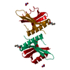
Crystal Structure of Leukocyte Ig-like Receptor A5 (LILRA5/LIR9/ILT11)
Components
leukocyte immunoglobulin-like receptor subfamily A member 5 isoform 1
Keywords
IMMUNE SYSTEM / Immunoglobulin-like fold
Function / homology
Function and homology information
positive regulation of cell activation / inhibitory MHC class I receptor activity / negative regulation of interleukin-13 production / immune response-regulating signaling pathway / positive regulation of protein tyrosine kinase activity / negative regulation of interleukin-12 production / positive regulation of calcium ion transport / positive regulation of interleukin-10 production / positive regulation of interleukin-1 beta production / positive regulation of interleukin-6 production ...positive regulation of cell activation / inhibitory MHC class I receptor activity / negative regulation of interleukin-13 production / immune response-regulating signaling pathway / positive regulation of protein tyrosine kinase activity / negative regulation of interleukin-12 production / positive regulation of calcium ion transport / positive regulation of interleukin-10 production / positive regulation of interleukin-1 beta production / positive regulation of interleukin-6 production / Immunoregulatory interactions between a Lymphoid and a non-Lymphoid cell / cytokine-mediated signaling pathway / positive regulation of tumor necrosis factor production / positive regulation of inflammatory response / positive regulation of MAPK cascade / innate immune response / cell surface / : / extracellular region / plasma membrane Similarity search - Function
Resolution: 1.8→50 Å / Num. obs: 19328 / % possible obs: 92.1 % / Observed criterion σ(I): 1 / Redundancy: 6.3 % / Rmerge(I) obs: 0.052 / Net I/σ(I): 16.6
Reflection shell
Resolution: 1.8→1.86 Å / Redundancy: 3.4 % / Rmerge(I) obs: 0.382 / Mean I/σ(I) obs: 2.5 / Num. unique all: 1011 / % possible all: 49.2
-
Processing
Software
Name
Version
Classification
REFMAC
5.2.0005
refinement
CNS
refinement
HKL-2000
datareduction
SCALEPACK
datascaling
SOLVE
phasing
Refinement
Method to determine structure: MAD / Resolution: 1.85→50 Å / Cor.coef. Fo:Fc: 0.934 / Cor.coef. Fo:Fc free: 0.908 / SU B: 3.364 / SU ML: 0.104 / Cross valid method: THROUGHOUT / σ(F): 0 / σ(I): 0 / ESU R: 0.163 / ESU R Free: 0.152 / Stereochemistry target values: MAXIMUM LIKELIHOOD Details: HYDROGENS HAVE BEEN ADDED IN THE RIDING POSITIONS. The structure was refined also with CNS.
Rfactor
Num. reflection
% reflection
Selection details
Rfree
0.26861
951
5.1 %
RANDOM
Rwork
0.23363
-
-
-
obs
0.23547
17645
95.62 %
-
Solvent computation
Ion probe radii: 0.8 Å / Shrinkage radii: 0.8 Å / VDW probe radii: 1.2 Å / Solvent model: MASK
Displacement parameters
Biso mean: 34.844 Å2
Baniso -1
Baniso -2
Baniso -3
1-
-1.17 Å2
0 Å2
0 Å2
2-
-
0.4 Å2
0 Å2
3-
-
-
0.76 Å2
Refinement step
Cycle: LAST / Resolution: 1.85→50 Å
Protein
Nucleic acid
Ligand
Solvent
Total
Num. atoms
1436
0
0
105
1541
Refine LS restraints
Refine-ID
Type
Dev ideal
Dev ideal target
Number
X-RAY DIFFRACTION
r_bond_refined_d
0.015
0.022
1481
X-RAY DIFFRACTION
r_bond_other_d
X-RAY DIFFRACTION
r_angle_refined_deg
1.551
1.961
2018
X-RAY DIFFRACTION
r_angle_other_deg
X-RAY DIFFRACTION
r_dihedral_angle_1_deg
7.272
5
176
X-RAY DIFFRACTION
r_dihedral_angle_2_deg
35.144
22.576
66
X-RAY DIFFRACTION
r_dihedral_angle_3_deg
18.481
15
238
X-RAY DIFFRACTION
r_dihedral_angle_4_deg
16.864
15
13
X-RAY DIFFRACTION
r_chiral_restr
0.102
0.2
215
X-RAY DIFFRACTION
r_gen_planes_refined
0.006
0.02
1139
X-RAY DIFFRACTION
r_gen_planes_other
X-RAY DIFFRACTION
r_nbd_refined
0.2
0.2
563
X-RAY DIFFRACTION
r_nbd_other
X-RAY DIFFRACTION
r_nbtor_refined
0.307
0.2
962
X-RAY DIFFRACTION
r_nbtor_other
X-RAY DIFFRACTION
r_xyhbond_nbd_refined
0.17
0.2
96
X-RAY DIFFRACTION
r_xyhbond_nbd_other
X-RAY DIFFRACTION
r_metal_ion_refined
X-RAY DIFFRACTION
r_metal_ion_other
X-RAY DIFFRACTION
r_symmetry_vdw_refined
0.169
0.2
37
X-RAY DIFFRACTION
r_symmetry_vdw_other
X-RAY DIFFRACTION
r_symmetry_hbond_refined
0.167
0.2
19
X-RAY DIFFRACTION
r_symmetry_hbond_other
X-RAY DIFFRACTION
r_symmetry_metal_ion_refined
X-RAY DIFFRACTION
r_symmetry_metal_ion_other
X-RAY DIFFRACTION
r_mcbond_it
1.046
1.5
922
X-RAY DIFFRACTION
r_mcbond_other
X-RAY DIFFRACTION
r_mcangle_it
1.701
2
1460
X-RAY DIFFRACTION
r_scbond_it
2.307
3
652
X-RAY DIFFRACTION
r_scangle_it
3.493
4.5
558
X-RAY DIFFRACTION
r_rigid_bond_restr
X-RAY DIFFRACTION
r_sphericity_free
X-RAY DIFFRACTION
r_sphericity_bonded
LS refinement shell
Resolution: 1.848→1.896 Å / Total num. of bins used: 20
Rfactor
Num. reflection
% reflection
Rfree
0.442
42
-
Rwork
0.297
910
-
obs
-
952
67.04 %
+
About Yorodumi
-
News
-
Feb 9, 2022. New format data for meta-information of EMDB entries
New format data for meta-information of EMDB entries
Version 3 of the EMDB header file is now the official format.
The previous official version 1.9 will be removed from the archive.
In the structure databanks used in Yorodumi, some data are registered as the other names, "COVID-19 virus" and "2019-nCoV". Here are the details of the virus and the list of structure data.
Jan 31, 2019. EMDB accession codes are about to change! (news from PDBe EMDB page)
EMDB accession codes are about to change! (news from PDBe EMDB page)
The allocation of 4 digits for EMDB accession codes will soon come to an end. Whilst these codes will remain in use, new EMDB accession codes will include an additional digit and will expand incrementally as the available range of codes is exhausted. The current 4-digit format prefixed with “EMD-” (i.e. EMD-XXXX) will advance to a 5-digit format (i.e. EMD-XXXXX), and so on. It is currently estimated that the 4-digit codes will be depleted around Spring 2019, at which point the 5-digit format will come into force.
The EM Navigator/Yorodumi systems omit the EMD- prefix.
Related info.:Q: What is EMD? / ID/Accession-code notation in Yorodumi/EM Navigator
Yorodumi is a browser for structure data from EMDB, PDB, SASBDB, etc.
This page is also the successor to EM Navigator detail page, and also detail information page/front-end page for Omokage search.
The word "yorodu" (or yorozu) is an old Japanese word meaning "ten thousand". "mi" (miru) is to see.
Related info.:EMDB / PDB / SASBDB / Comparison of 3 databanks / Yorodumi Search / Aug 31, 2016. New EM Navigator & Yorodumi / Yorodumi Papers / Jmol/JSmol / Function and homology information / Changes in new EM Navigator and Yorodumi
 Movie
Movie Controller

 Yorodumi
 Open data
 Basic information
 Components
 Keywords
 Function and homology information
 Homo sapiens (human)
 X-RAY DIFFRACTION /
  Authors
 Citation
 Structure visualization
 Downloads & links
 Other downloads
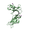


 PDBj
PDBj
 Assembly

 Mass: 18.015 Da / Num. of mol.: 105 / Source method: isolated from a natural source / Formula: H2O
 Sample preparation
 / Beamline: BL41XU / Wavelength: 0.97921, 0.97956, 0.90000, 1.0000
 Processing