| 登録情報 | データベース: PDB / ID: 2g56
|
|---|
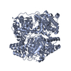
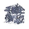
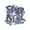
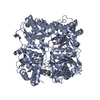
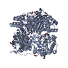
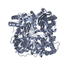
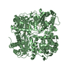
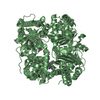
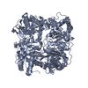
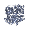
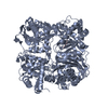
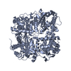
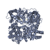
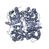
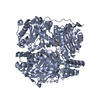
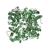
| タイトル | crystal structure of human insulin-degrading enzyme in complex with insulin B chain |
|---|
 要素 要素 | - Insulin
- Insulin-degrading enzyme
|
|---|
 キーワード キーワード | HYDROLASE / protein-peptide complex |
|---|
| 機能・相同性 |  機能・相同性情報 機能・相同性情報
insulysin / beta-endorphin binding / ubiquitin recycling / insulin catabolic process / insulin metabolic process / amyloid-beta clearance by cellular catabolic process / hormone catabolic process / bradykinin catabolic process / cytosolic proteasome complex / positive regulation of protein binding ...insulysin / beta-endorphin binding / ubiquitin recycling / insulin catabolic process / insulin metabolic process / amyloid-beta clearance by cellular catabolic process / hormone catabolic process / bradykinin catabolic process / cytosolic proteasome complex / positive regulation of protein binding / insulin binding / negative regulation of glycogen catabolic process / : / regulation of aerobic respiration / negative regulation of fatty acid metabolic process / Signaling by Insulin receptor / negative regulation of feeding behavior / IRS activation / peptide catabolic process / Insulin processing / regulation of protein secretion / positive regulation of peptide hormone secretion / positive regulation of respiratory burst / negative regulation of acute inflammatory response / Regulation of gene expression in beta cells / peroxisomal matrix / alpha-beta T cell activation / amyloid-beta clearance / Synthesis, secretion, and deacylation of Ghrelin / positive regulation of dendritic spine maintenance / amyloid-beta metabolic process / negative regulation of protein secretion / negative regulation of gluconeogenesis / fatty acid homeostasis / positive regulation of glycogen biosynthetic process / positive regulation of insulin receptor signaling pathway / Signal attenuation / FOXO-mediated transcription of oxidative stress, metabolic and neuronal genes / negative regulation of respiratory burst involved in inflammatory response / negative regulation of lipid catabolic process / positive regulation of lipid biosynthetic process / negative regulation of oxidative stress-induced intrinsic apoptotic signaling pathway / nitric oxide-cGMP-mediated signaling / regulation of protein localization to plasma membrane / transport vesicle / positive regulation of nitric-oxide synthase activity / Insulin receptor recycling / COPI-mediated anterograde transport / negative regulation of reactive oxygen species biosynthetic process / positive regulation of brown fat cell differentiation / insulin-like growth factor receptor binding / negative regulation of proteolysis / NPAS4 regulates expression of target genes / peptide binding / neuron projection maintenance / endoplasmic reticulum-Golgi intermediate compartment membrane / positive regulation of mitotic nuclear division / Insulin receptor signalling cascade / : / positive regulation of glycolytic process / endosome lumen / acute-phase response / positive regulation of cytokine production / positive regulation of long-term synaptic potentiation / positive regulation of D-glucose import across plasma membrane / positive regulation of protein secretion / insulin receptor binding / protein catabolic process / positive regulation of cell differentiation / wound healing / Regulation of insulin secretion / Peroxisomal protein import / hormone activity / antigen processing and presentation of endogenous peptide antigen via MHC class I / positive regulation of neuron projection development / metalloendopeptidase activity / negative regulation of protein catabolic process / regulation of synaptic plasticity / positive regulation of protein localization to nucleus / Golgi lumen / vasodilation / cognition / glucose metabolic process / positive regulation of protein catabolic process / insulin receptor signaling pathway / peroxisome / cell-cell signaling / regulation of protein localization / glucose homeostasis / amyloid-beta binding / PI5P, PP2A and IER3 Regulate PI3K/AKT Signaling / virus receptor activity / positive regulation of cell growth / protease binding / secretory granule lumen / endopeptidase activity / basolateral plasma membrane / positive regulation of canonical NF-kappaB signal transduction / positive regulation of MAPK cascade / positive regulation of phosphatidylinositol 3-kinase/protein kinase B signal transduction類似検索 - 分子機能 Peptidase M16, middle/third domain / Middle or third domain of peptidase_M16 / : / PQQ synthase PqqF-like, C-terminal lobe domain 4 / : / Cytochrome Bc1 Complex; Chain A, domain 1 / Metalloenzyme, LuxS/M16 peptidase-like / Peptidase M16, zinc-binding site / Insulinase family, zinc-binding region signature. / Peptidase M16, C-terminal ...Peptidase M16, middle/third domain / Middle or third domain of peptidase_M16 / : / PQQ synthase PqqF-like, C-terminal lobe domain 4 / : / Cytochrome Bc1 Complex; Chain A, domain 1 / Metalloenzyme, LuxS/M16 peptidase-like / Peptidase M16, zinc-binding site / Insulinase family, zinc-binding region signature. / Peptidase M16, C-terminal / Peptidase M16 inactive domain / Peptidase M16, N-terminal / Insulinase (Peptidase family M16) / Metalloenzyme, LuxS/M16 peptidase-like / Insulin / Insulin family / Insulin-like / Insulin/IGF/Relaxin family / Insulin / insulin-like growth factor / relaxin family. / Insulin, conserved site / Insulin family signature. / Insulin-like superfamily / 2-Layer Sandwich / Alpha Beta類似検索 - ドメイン・相同性 1,4-DIETHYLENE DIOXIDE / Insulin / Insulin-degrading enzyme / Insulin-degrading enzyme類似検索 - 構成要素 |
|---|
| 生物種 |  Homo sapiens (ヒト) Homo sapiens (ヒト) |
|---|
| 手法 |  X線回折 / シンクロトロン / 分子置換 / 解像度: 2.2 Å X線回折 / シンクロトロン / 分子置換 / 解像度: 2.2 Å |
|---|
 データ登録者 データ登録者 | Shen, Y. / Tang, W.-J. |
|---|
 引用 引用 | ジャーナル: Nature / 年: 2006
タイトル: Structures of human insulin-degrading enzyme reveal a new substrate recognition mechanism.
著者: Shen, Y. / Joachimiak, A. / Rosner, M.R. / Tang, W.J. |
|---|
| 履歴 | | 登録 | 2006年2月22日 | 登録サイト: RCSB / 処理サイト: RCSB |
|---|
| 改定 1.0 | 2006年10月24日 | Provider: repository / タイプ: Initial release |
|---|
| 改定 1.1 | 2008年5月1日 | Group: Version format compliance |
|---|
| 改定 1.2 | 2011年7月13日 | Group: Version format compliance |
|---|
| 改定 1.3 | 2021年10月20日 | Group: Database references / Derived calculations / カテゴリ: database_2 / struct_ref_seq_dif / struct_site
Item: _database_2.pdbx_DOI / _database_2.pdbx_database_accession ..._database_2.pdbx_DOI / _database_2.pdbx_database_accession / _struct_ref_seq_dif.details / _struct_site.pdbx_auth_asym_id / _struct_site.pdbx_auth_comp_id / _struct_site.pdbx_auth_seq_id |
|---|
| 改定 1.4 | 2023年8月30日 | Group: Data collection / Refinement description
カテゴリ: chem_comp_atom / chem_comp_bond / pdbx_initial_refinement_model |
|---|
|
|---|
 ムービー
ムービー コントローラー


 データを開く
 基本情報
基本情報 構造の表示
構造の表示 ダウンロードとリンク
 その他のダウンロード


 PDBj
PDBj


 集合体
集合体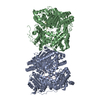


 分子量: 88.105 Da / 分子数: 1 / 由来タイプ: 合成 / 式: C4H8O2
 分子量: 18.015 Da / 分子数: 1044 / 由来タイプ: 天然 / 式: H2O
 試料調製
試料調製 / ビームライン: 19-ID / 波長: 0.9 Å
 解析
解析