 Movie
Movie Controller

[English] 日本語
 Yorodumi
- PDB-2fec: Structure of the E203Q mutant of the Cl-/H+ exchanger CLC-ec1 fro... -
+ Open data

- Basic information
| Entry | Database: PDB / ID: 2fec | ||||||
|---|---|---|---|---|---|---|---|
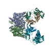
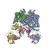
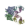
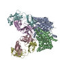
| Title | Structure of the E203Q mutant of the Cl-/H+ exchanger CLC-ec1 from E.Coli | ||||||
 Components Components |
| ||||||
 Keywords Keywords | PROTON TRANSPORT / MEMBRANE PROTEIN / CLC-ec1 / CLCA_ECOLI / Chloride/Proton exchange transporter | ||||||
| Function / homology |  Function and homology information Function and homology informationcellular stress response to acidic pH / chloride:proton antiporter activity / voltage-gated chloride channel activity / proton transmembrane transport / chloride transmembrane transport / identical protein binding / plasma membrane Similarity search - Function | ||||||
| Biological species |   Homo sapiens (human) Homo sapiens (human) | ||||||
| Method |  X-RAY DIFFRACTION / SYNCHROTRON / MOLECULAR REPLACEMENT / Resolution: 3.967 Å X-RAY DIFFRACTION / SYNCHROTRON / MOLECULAR REPLACEMENT / Resolution: 3.967 Å | ||||||
 Authors Authors | Accardi, A. / Walden, M.P. / Nguitragool, W. / Jayaram, H. / Williams, C. / Miller, C. | ||||||
 Citation Citation | Journal: J.Gen.Physiol. / Year: 2005 Title: Separate ion pathways in a Cl-/H+ exchanger Authors: Accardi, A. / Walden, M.P. / Nguitragool, W. / Jayaram, H. / Williams, C. / Miller, C. #1: Journal: Science / Year: 2003Title: Gating the selectivity filter in ClC chloride channels Authors: Dutzler, R. / Cambell, E.B. / Mackinnon, R. | ||||||
| History |
|
- Structure visualization
| Structure viewer | Molecule: MolmilJmol/JSmol |
|---|
- Downloads & links
-Download
| PDBx/mmCIF format | 2fec.cif.gz | 325 KB | Display | PDBx/mmCIF format |
|---|---|---|---|---|
| PDB format | pdb2fec.ent.gz | 265.4 KB | Display | PDB format |
| PDBx/mmJSON format | 2fec.json.gz | Tree view | PDBx/mmJSON format | |
| Others |  Other downloads Other downloads |
-Validation report
| Arichive directory | https://data.pdbj.org/pub/pdb/validation_reports/fe/2fecftp://data.pdbj.org/pub/pdb/validation_reports/fe/2fec | HTTPS FTP |
|---|
-Related structure data


-Links
 PDBj
PDBj

- Assembly
| Deposited unit | 
| ||||||||||||||||||
|---|---|---|---|---|---|---|---|---|---|---|---|---|---|---|---|---|---|---|---|
| 1 |
| ||||||||||||||||||
| Unit cell |
| ||||||||||||||||||
| Noncrystallographic symmetry (NCS) | NCS domain:
NCS domain segments: Component-ID: 1 / Ens-ID: 1 / Beg auth comp-ID: ARG / Beg label comp-ID: ARG / End auth comp-ID: ALA / End label comp-ID: ALA / Refine code: 3 / Auth seq-ID: 18 - 458 / Label seq-ID: 18 - 458
|
-Components
| #1: Protein | Mass: 49657.711 Da / Num. of mol.: 2 / Mutation: E203Q Source method: isolated from a genetically manipulated source Source: (gene. exp.) #2: Antibody | Mass: 23823.031 Da / Num. of mol.: 2 / Fragment: Heavy Chain Source method: isolated from a genetically manipulated source Source: (gene. exp.) Homo sapiens (human) / Cell (production host): 10EC3/G4 / Cell line (production host): hybridoma / Production host:  #3: Antibody | Mass: 23088.443 Da / Num. of mol.: 2 / Fragment: Light Chain Source method: isolated from a genetically manipulated source Source: (gene. exp.) Homo sapiens (human) / Cell (production host): 10EC3/G4 / Cell line (production host): hybridoma / Production host: Has protein modification | Y | |
|---|
-Experimental details
-Experiment
| Experiment | Method: X-RAY DIFFRACTION / Number of used crystals: 1 |
|---|
- Sample preparation
| Crystal | Density Matthews: 3.73 Å3/Da / Density % sol: 67.05 % |
|---|---|
| Crystal grow | Temperature: 298 K / Method: vapor diffusion, sitting drop / pH: 7.5 Details: 250 mM NaBr, PEG 400 37%, 0.05 M Hepes, pH 7.5, VAPOR DIFFUSION, SITTING DROP, temperature 298K |
-Data collection
| Diffraction | Mean temperature: 100 K |
|---|---|
| Diffraction source | Source: SYNCHROTRON / Site: APS  / Beamline: 19-ID / Wavelength: 0.9198 Å / Beamline: 19-ID / Wavelength: 0.9198 Å |
| Detector | Detector: CCD |
| Radiation | Protocol: SINGLE WAVELENGTH / Monochromatic (M) / Laue (L): M / Scattering type: x-ray |
| Radiation wavelength | Wavelength: 0.9198 Å / Relative weight: 1 |
| Reflection | Resolution: 3.967→128.04 Å / Num. all: 25143 / Num. obs: 23320 / % possible obs: 98.5 % / Observed criterion σ(I): 2 / Rmerge(I) obs: 0.085 / Rsym value: 0.085 / Net I/σ(I): 13.3 |
- Processing
| Software |
| ||||||||||||||||||||||||||||||||||||||||||||||||||||||||||||||||||||||||||||||||||||||||||||||||||||||||||||||||||||||||||||||||||||||||||||||||||||||||||||||||||||||||||
|---|---|---|---|---|---|---|---|---|---|---|---|---|---|---|---|---|---|---|---|---|---|---|---|---|---|---|---|---|---|---|---|---|---|---|---|---|---|---|---|---|---|---|---|---|---|---|---|---|---|---|---|---|---|---|---|---|---|---|---|---|---|---|---|---|---|---|---|---|---|---|---|---|---|---|---|---|---|---|---|---|---|---|---|---|---|---|---|---|---|---|---|---|---|---|---|---|---|---|---|---|---|---|---|---|---|---|---|---|---|---|---|---|---|---|---|---|---|---|---|---|---|---|---|---|---|---|---|---|---|---|---|---|---|---|---|---|---|---|---|---|---|---|---|---|---|---|---|---|---|---|---|---|---|---|---|---|---|---|---|---|---|---|---|---|---|---|---|---|---|---|---|
| Refinement | Method to determine structure: MOLECULAR REPLACEMENT / Resolution: 3.967→128.04 Å / Cor.coef. Fo:Fc: 0.896 / Cor.coef. Fo:Fc free: 0.866 / SU B: 59.536 / SU ML: 0.826 / Cross valid method: THROUGHOUT / σ(F): 3 / ESU R Free: 0.997 / Stereochemistry target values: MAXIMUM LIKELIHOOD / Details: HYDROGENS HAVE BEEN ADDED IN THE RIDING POSITIONS
| ||||||||||||||||||||||||||||||||||||||||||||||||||||||||||||||||||||||||||||||||||||||||||||||||||||||||||||||||||||||||||||||||||||||||||||||||||||||||||||||||||||||||||
| Solvent computation | Ion probe radii: 0.8 Å / Shrinkage radii: 0.8 Å / VDW probe radii: 1.4 Å / Solvent model: MASK | ||||||||||||||||||||||||||||||||||||||||||||||||||||||||||||||||||||||||||||||||||||||||||||||||||||||||||||||||||||||||||||||||||||||||||||||||||||||||||||||||||||||||||
| Displacement parameters | Biso mean: 155.801 Å2
| ||||||||||||||||||||||||||||||||||||||||||||||||||||||||||||||||||||||||||||||||||||||||||||||||||||||||||||||||||||||||||||||||||||||||||||||||||||||||||||||||||||||||||
| Refinement step | Cycle: LAST / Resolution: 3.967→128.04 Å
| ||||||||||||||||||||||||||||||||||||||||||||||||||||||||||||||||||||||||||||||||||||||||||||||||||||||||||||||||||||||||||||||||||||||||||||||||||||||||||||||||||||||||||
| Refine LS restraints |
| ||||||||||||||||||||||||||||||||||||||||||||||||||||||||||||||||||||||||||||||||||||||||||||||||||||||||||||||||||||||||||||||||||||||||||||||||||||||||||||||||||||||||||
| Refine LS restraints NCS | Dom-ID: 1 / Auth asym-ID: A / Ens-ID: 1 / Refine-ID: X-RAY DIFFRACTION
| ||||||||||||||||||||||||||||||||||||||||||||||||||||||||||||||||||||||||||||||||||||||||||||||||||||||||||||||||||||||||||||||||||||||||||||||||||||||||||||||||||||||||||
| LS refinement shell | Resolution: 3.967→4.069 Å / Total num. of bins used: 20
|
