 Movie
Movie Controller

[English] 日本語
 Yorodumi
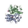
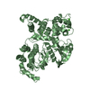
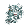
- PDB-2f8d: BenM effector-Binding domain crystallized from high pH conditions -
+ Open data

- Basic information
| Entry | Database: PDB / ID: 2f8d | ||||||
|---|---|---|---|---|---|---|---|
| Title | BenM effector-Binding domain crystallized from high pH conditions | ||||||
 Components Components | HTH-type transcriptional regulator benM | ||||||
 Keywords Keywords | GENE REGULATION / BenM / LTTR / LysR-type transcriptional regulator / tetramerization / Effector binding domain / Inducer binding domain | ||||||
| Function / homology |  Function and homology information Function and homology informationprotein-DNA complex / DNA-binding transcription factor activity / DNA binding Similarity search - Function | ||||||
| Biological species |  Acinetobacter baylyi (bacteria) Acinetobacter baylyi (bacteria) | ||||||
| Method |  X-RAY DIFFRACTION / SYNCHROTRON / MOLECULAR REPLACEMENT / Resolution: 2.7 Å X-RAY DIFFRACTION / SYNCHROTRON / MOLECULAR REPLACEMENT / Resolution: 2.7 Å | ||||||
 Authors Authors | Ezezika, O.C. / Haddad, S. / Neidle, E.L. / Momany, C. | ||||||
 Citation Citation | Journal: Acta Crystallogr.,Sect.F / Year: 2007 Title: Oligomerization of BenM, a LysR-type transcriptional regulator: structural basis for the aggregation of proteins in this family. Authors: Ezezika, O.C. / Haddad, S. / Neidle, E.L. / Momany, C. #1: Journal: Acta Crystallogr.,Sect.D / Year: 2004Title: Crystallization of the effector-binding domains of BenM and BenM, LysR-type transcriptional regulators from Acinetobacter sp. ADP1. Authors: Clark, T. / Haddad, S. / Neidle, E. / Momany, C. | ||||||
| History |
|
- Structure visualization
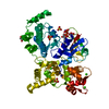
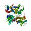
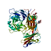
| Structure viewer | Molecule: MolmilJmol/JSmol |
|---|
- Downloads & links
-Download
| PDBx/mmCIF format | 2f8d.cif.gz | 112.5 KB | Display | PDBx/mmCIF format |
|---|---|---|---|---|
| PDB format | pdb2f8d.ent.gz | 86.4 KB | Display | PDB format |
| PDBx/mmJSON format | 2f8d.json.gz | Tree view | PDBx/mmJSON format | |
| Others |  Other downloads Other downloads |
-Validation report
| Arichive directory | https://data.pdbj.org/pub/pdb/validation_reports/f8/2f8dftp://data.pdbj.org/pub/pdb/validation_reports/f8/2f8d | HTTPS FTP |
|---|
-Related structure data
| Related structure data |  2f97C  2f6gS S: Starting model for refinement C: citing same article ( |
|---|---|
| Similar structure data |


-Links
 PDBj
PDBj- Assembly
| Deposited unit | 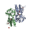
| ||||||||
|---|---|---|---|---|---|---|---|---|---|
| 1 |
| ||||||||
| Unit cell |
| ||||||||
| Components on special symmetry positions |
| ||||||||
| Details | The biological unit is a dimer. A dimer is in the asymmetric unit. |
-Components
| #1: Protein | Mass: 26401.447 Da / Num. of mol.: 2 Source method: isolated from a genetically manipulated source Source: (gene. exp.) Acinetobacter baylyi (bacteria) / Strain: ADP1 / Gene: benM / Plasmid: pet21b / Production host: #2: Chemical |   Mass: 122.121 Da / Num. of mol.: 3 / Source method: obtained synthetically / Formula: C7H6O2 Mass: 122.121 Da / Num. of mol.: 3 / Source method: obtained synthetically / Formula: C7H6O2#3: Chemical | ChemComp-GOL /   Mass: 92.094 Da / Num. of mol.: 6 / Source method: obtained synthetically / Formula: C3H8O3 Mass: 92.094 Da / Num. of mol.: 6 / Source method: obtained synthetically / Formula: C3H8O3#4: Chemical | ChemComp-PO4 / |   Mass: 94.971 Da / Num. of mol.: 1 / Source method: obtained synthetically / Formula: PO4 Mass: 94.971 Da / Num. of mol.: 1 / Source method: obtained synthetically / Formula: PO4#5: Water | ChemComp-HOH / |  Mass: 18.015 Da / Num. of mol.: 455 / Source method: isolated from a natural source / Formula: H2O Mass: 18.015 Da / Num. of mol.: 455 / Source method: isolated from a natural source / Formula: H2O |
|---|
-Experimental details
-Experiment
| Experiment | Method: X-RAY DIFFRACTION / Number of used crystals: 1 |
|---|
- Sample preparation
| Crystal | Density Matthews: 4.6 Å3/Da / Density % sol: 73.1 % |
|---|---|
| Crystal grow | pH: 10 Details: PEG 4000, glycerol, imidazole, tris, CAPS, KBr, NaCl, benzoate, pH 10, Microbatch under oil |
-Data collection
| Diffraction | Mean temperature: 100 K |
|---|---|
| Diffraction source | Source: SYNCHROTRON / Site: APS  / Beamline: 19-BM / Wavelength: 0.97935 Å / Beamline: 19-BM / Wavelength: 0.97935 Å |
| Detector | Type: MARRESEARCH / Detector: CCD / Date: Jan 1, 2003 |
| Radiation | Protocol: SINGLE WAVELENGTH / Monochromatic (M) / Laue (L): M / Scattering type: x-ray |
| Radiation wavelength | Wavelength: 0.97935 Å / Relative weight: 1 |
| Reflection | Resolution: 2.7→92.06 Å / Num. all: 25480 / Num. obs: 25480 / % possible obs: 98.4 % / Observed criterion σ(F): 0 / Observed criterion σ(I): 0 / Redundancy: 7.16 % / Biso Wilson estimate: 53.78698 Å2 / Rmerge(I) obs: 0.093 / Net I/σ(I): 28.1 |
| Reflection shell | Resolution: 2.7→2.8 Å / % possible obs: 98.1 % / Rmerge(I) obs: 0.421 / Mean I/σ(I) obs: 5.85 / Num. measured obs: 2494 / Num. unique all: 2494 / Rsym value: 0 / Χ2: 1.355 / % possible all: 98.1 |
- Processing
| Software |
| ||||||||||||||||||||||||||||||||||||||||||||||||||||||||||||||||||||||||||||||||||||||||||
|---|---|---|---|---|---|---|---|---|---|---|---|---|---|---|---|---|---|---|---|---|---|---|---|---|---|---|---|---|---|---|---|---|---|---|---|---|---|---|---|---|---|---|---|---|---|---|---|---|---|---|---|---|---|---|---|---|---|---|---|---|---|---|---|---|---|---|---|---|---|---|---|---|---|---|---|---|---|---|---|---|---|---|---|---|---|---|---|---|---|---|---|
| Refinement | Method to determine structure: MOLECULAR REPLACEMENT Starting model: PDB ENTRY 2F6G Resolution: 2.7→92.06 Å / Cor.coef. Fo:Fc: 0.961 / Cor.coef. Fo:Fc free: 0.933 / SU B: 12.356 / SU ML: 0.139 / Isotropic thermal model: TLS / Cross valid method: THROUGHOUT / σ(F): 0 / ESU R: 0.291 / ESU R Free: 0.221 / Stereochemistry target values: Engh & Huber / Details: HYDROGENS HAVE BEEN ADDED IN THE RIDING POSITIONS
| ||||||||||||||||||||||||||||||||||||||||||||||||||||||||||||||||||||||||||||||||||||||||||
| Solvent computation | Ion probe radii: 0.8 Å / Shrinkage radii: 0.8 Å / VDW probe radii: 1.2 Å / Solvent model: BABINET MODEL WITH MASK | ||||||||||||||||||||||||||||||||||||||||||||||||||||||||||||||||||||||||||||||||||||||||||
| Displacement parameters | Biso mean: 32.036 Å2
| ||||||||||||||||||||||||||||||||||||||||||||||||||||||||||||||||||||||||||||||||||||||||||
| Refinement step | Cycle: LAST / Resolution: 2.7→92.06 Å
| ||||||||||||||||||||||||||||||||||||||||||||||||||||||||||||||||||||||||||||||||||||||||||
| Refine LS restraints |
| ||||||||||||||||||||||||||||||||||||||||||||||||||||||||||||||||||||||||||||||||||||||||||
| LS refinement shell | Resolution: 2.695→2.765 Å / Total num. of bins used: 20
|
