- PDB-4k84: Crystal structure of human ceramide-1-phosphate transfer protein ... -
+
Open data
ID or keywords:
Loading...
-
Basic information
Entry
Database: PDB / ID: 4k84
Title
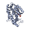
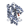
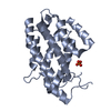
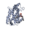
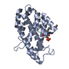
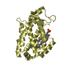
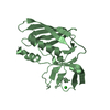
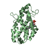
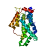
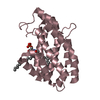
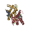
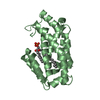
Crystal structure of human ceramide-1-phosphate transfer protein (CPTP) in complex with 16:0 ceramide-1-phosphate (16:0-C1P)
Components
Glycolipid transfer protein domain-containing protein 1
Keywords
LIPID TRANSPORT / Lipid transfer protein / GLTP-fold / CPTP / C1P / Ceramide-1-phosphate / Protein-lipid complex / Eicosanoid
Function / homology
Function and homology information
ceramide 1-phosphate transport / ceramide 1-phosphate transfer activity / ceramide transport / ceramide 1-phosphate binding / Glycosphingolipid transport / intermembrane lipid transfer / negative regulation of NLRP3 inflammasome complex assembly / negative regulation of interleukin-1 beta production / perinuclear theca / nuclear outer membrane ...ceramide 1-phosphate transport / ceramide 1-phosphate transfer activity / ceramide transport / ceramide 1-phosphate binding / Glycosphingolipid transport / intermembrane lipid transfer / negative regulation of NLRP3 inflammasome complex assembly / negative regulation of interleukin-1 beta production / perinuclear theca / nuclear outer membrane / negative regulation of autophagy / centriole / phospholipid binding / sperm midpiece / endosome membrane / Golgi apparatus / plasma membrane / cytosol Similarity search - Function
Glycolipid transfer protein domain / Glycolipid transfer protein superfamily / Glycolipid transfer protein (GLTP) / Glycolipid transfer protein, GLTP / Glycolipid transfer protein / Orthogonal Bundle / Mainly Alpha Similarity search - Domain/homology
In the structure databanks used in Yorodumi, some data are registered as the other names, "COVID-19 virus" and "2019-nCoV". Here are the details of the virus and the list of structure data.
Jan 31, 2019. EMDB accession codes are about to change! (news from PDBe EMDB page)
EMDB accession codes are about to change! (news from PDBe EMDB page)
The allocation of 4 digits for EMDB accession codes will soon come to an end. Whilst these codes will remain in use, new EMDB accession codes will include an additional digit and will expand incrementally as the available range of codes is exhausted. The current 4-digit format prefixed with “EMD-” (i.e. EMD-XXXX) will advance to a 5-digit format (i.e. EMD-XXXXX), and so on. It is currently estimated that the 4-digit codes will be depleted around Spring 2019, at which point the 5-digit format will come into force.
The EM Navigator/Yorodumi systems omit the EMD- prefix.
Related info.:Q: What is EMD? / ID/Accession-code notation in Yorodumi/EM Navigator
Yorodumi is a browser for structure data from EMDB, PDB, SASBDB, etc.
This page is also the successor to EM Navigator detail page, and also detail information page/front-end page for Omokage search.
The word "yorodu" (or yorozu) is an old Japanese word meaning "ten thousand". "mi" (miru) is to see.
Related info.:EMDB / PDB / SASBDB / Comparison of 3 databanks / Yorodumi Search / Aug 31, 2016. New EM Navigator & Yorodumi / Yorodumi Papers / Jmol/JSmol / Function and homology information / Changes in new EM Navigator and Yorodumi
 Movie
Movie Controller

 Yorodumi
 Open data
 Basic information
 Components
 Keywords
 Function and homology information
 Homo sapiens (human)
 X-RAY DIFFRACTION /
  Authors
 Citation
 Structure visualization
 Downloads & links
 Other downloads
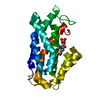


 PDBj
PDBj Assembly


 Mass: 18.015 Da / Num. of mol.: 439 / Source method: isolated from a natural source / Formula: H2O
 Sample preparation
 / Beamline: 24-ID-C / Wavelength: 1.2398 Å
 Processing