 Movie
Movie Controller

[English] 日本語
 Yorodumi
- PDB-4kbr: Crystal structure of mouse Ceramide-1-phosphate transfer protein ... -
+ Open data

- Basic information
| Entry | Database: PDB / ID: 4kbr | ||||||
|---|---|---|---|---|---|---|---|
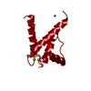
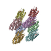
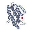
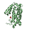
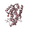
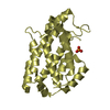
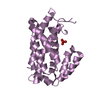
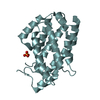
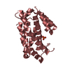
| Title | Crystal structure of mouse Ceramide-1-phosphate transfer protein (apo-form) | ||||||
 Components Components | Glycolipid transfer protein domain-containing protein 1 | ||||||
 Keywords Keywords | LIPID TRANSPORT / Lipid transfer protein / GLTP-fold / CPTP / C1P / Ceramide-1-phosphate / Protein-lipid complex / Eicosanoid | ||||||
| Function / homology |  Function and homology information Function and homology informationGlycosphingolipid transport / ceramide 1-phosphate transport / ceramide 1-phosphate transfer activity / ceramide 1-phosphate binding / negative regulation of NLRP3 inflammasome complex assembly / negative regulation of interleukin-1 beta production / nuclear outer membrane / negative regulation of autophagy / phospholipid binding / endosome membrane ...Glycosphingolipid transport / ceramide 1-phosphate transport / ceramide 1-phosphate transfer activity / ceramide 1-phosphate binding / negative regulation of NLRP3 inflammasome complex assembly / negative regulation of interleukin-1 beta production / nuclear outer membrane / negative regulation of autophagy / phospholipid binding / endosome membrane / Golgi apparatus / plasma membrane / cytosol Similarity search - Function | ||||||
| Biological species |  | ||||||
| Method |  X-RAY DIFFRACTION / SYNCHROTRON / MOLECULAR REPLACEMENT / molecular replacement / Resolution: 2.547 Å X-RAY DIFFRACTION / SYNCHROTRON / MOLECULAR REPLACEMENT / molecular replacement / Resolution: 2.547 Å | ||||||
 Authors Authors | Simanshu, D.K. / Brown, R.E. / Patel, D.J. | ||||||
 Citation Citation | Journal: Nature / Year: 2013 Title: Non-vesicular trafficking by a ceramide-1-phosphate transfer protein regulates eicosanoids. Authors: Simanshu, D.K. / Kamlekar, R.K. / Wijesinghe, D.S. / Zou, X. / Zhai, X. / Mishra, S.K. / Molotkovsky, J.G. / Malinina, L. / Hinchcliffe, E.H. / Chalfant, C.E. / Brown, R.E. / Patel, D.J. | ||||||
| History |
|
- Structure visualization
| Structure viewer | Molecule: MolmilJmol/JSmol |
|---|
- Downloads & links
-Download
| PDBx/mmCIF format | 4kbr.cif.gz | 640 KB | Display | PDBx/mmCIF format |
|---|---|---|---|---|
| PDB format | pdb4kbr.ent.gz | 535.1 KB | Display | PDB format |
| PDBx/mmJSON format | 4kbr.json.gz | Tree view | PDBx/mmJSON format | |
| Others |  Other downloads Other downloads |
-Validation report
| Arichive directory | https://data.pdbj.org/pub/pdb/validation_reports/kb/4kbrftp://data.pdbj.org/pub/pdb/validation_reports/kb/4kbr | HTTPS FTP |
|---|
-Related structure data
| Related structure data | 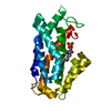 4k80C 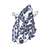 4k84C 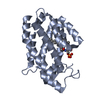 4k85C 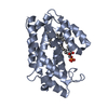 4k8nC 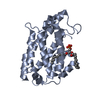 4kbsC  4kf6C C: citing same article ( |
|---|---|
| Similar structure data |


-Links
 PDBj
PDBj- Assembly

-Components
| #1: Protein | Mass: 24747.330 Da / Num. of mol.: 8 Source method: isolated from a genetically manipulated source Source: (gene. exp.)  #2: Chemical | ChemComp-PO4 /   Mass: 94.971 Da / Num. of mol.: 8 / Source method: obtained synthetically / Formula: PO4 Mass: 94.971 Da / Num. of mol.: 8 / Source method: obtained synthetically / Formula: PO4#3: Water | ChemComp-HOH / |  Mass: 18.015 Da / Num. of mol.: 254 / Source method: isolated from a natural source / Formula: H2O Mass: 18.015 Da / Num. of mol.: 254 / Source method: isolated from a natural source / Formula: H2O |
|---|
-Experimental details
-Experiment
| Experiment | Method: X-RAY DIFFRACTION / Number of used crystals: 1 |
|---|
- Sample preparation
| Crystal | Density Matthews: 2.33 Å3/Da / Density % sol: 47.3 % |
|---|---|
| Crystal grow | Temperature: 293 K / Method: vapor diffusion, sitting drop / pH: 6.5 Details: 0.1 M Bis-Tris pH 6.5, 2 M Ammonium sulfate, VAPOR DIFFUSION, SITTING DROP, temperature 293K |
-Data collection
| Diffraction | Mean temperature: 100 K | |||||||||||||||||||||||||||||||||||||||||||||||||||||||||||||||||||||||||||||
|---|---|---|---|---|---|---|---|---|---|---|---|---|---|---|---|---|---|---|---|---|---|---|---|---|---|---|---|---|---|---|---|---|---|---|---|---|---|---|---|---|---|---|---|---|---|---|---|---|---|---|---|---|---|---|---|---|---|---|---|---|---|---|---|---|---|---|---|---|---|---|---|---|---|---|---|---|---|---|
| Diffraction source | Source: SYNCHROTRON / Site: APS  / Beamline: 24-ID-E / Wavelength: 0.97918 Å / Beamline: 24-ID-E / Wavelength: 0.97918 Å | |||||||||||||||||||||||||||||||||||||||||||||||||||||||||||||||||||||||||||||
| Detector | Type: ADSC QUANTUM 315r / Detector: CCD / Date: Feb 1, 2010 | |||||||||||||||||||||||||||||||||||||||||||||||||||||||||||||||||||||||||||||
| Radiation | Monochromator: Si(111) / Protocol: SINGLE WAVELENGTH / Monochromatic (M) / Laue (L): M / Scattering type: x-ray | |||||||||||||||||||||||||||||||||||||||||||||||||||||||||||||||||||||||||||||
| Radiation wavelength | Wavelength: 0.97918 Å / Relative weight: 1 | |||||||||||||||||||||||||||||||||||||||||||||||||||||||||||||||||||||||||||||
| Reflection | Resolution: 2.55→50 Å / Num. obs: 57653 / % possible obs: 93.5 % / Redundancy: 10 % / Rmerge(I) obs: 0.086 / Χ2: 1.091 / Net I/σ(I): 8.3 | |||||||||||||||||||||||||||||||||||||||||||||||||||||||||||||||||||||||||||||
| Reflection shell |
|
-Phasing
| Phasing | Method: molecular replacement |
|---|
- Processing
| Software |
| ||||||||||||||||||||||||||||||||||||||||||||||||||||||||||||||||||||||||||||||||||||||||||||||||||||||||||||||||||||||||||||||||||||||||||||||||||||||||||
|---|---|---|---|---|---|---|---|---|---|---|---|---|---|---|---|---|---|---|---|---|---|---|---|---|---|---|---|---|---|---|---|---|---|---|---|---|---|---|---|---|---|---|---|---|---|---|---|---|---|---|---|---|---|---|---|---|---|---|---|---|---|---|---|---|---|---|---|---|---|---|---|---|---|---|---|---|---|---|---|---|---|---|---|---|---|---|---|---|---|---|---|---|---|---|---|---|---|---|---|---|---|---|---|---|---|---|---|---|---|---|---|---|---|---|---|---|---|---|---|---|---|---|---|---|---|---|---|---|---|---|---|---|---|---|---|---|---|---|---|---|---|---|---|---|---|---|---|---|---|---|---|---|---|---|---|
| Refinement | Method to determine structure: MOLECULAR REPLACEMENT Starting model: Human CPTP in complex with 2:0 C1P Resolution: 2.547→39.507 Å / Occupancy max: 1 / Occupancy min: 1 / SU ML: 0.39 / Cross valid method: THROUGHOUT / σ(F): 1.34 / Phase error: 31.09 / Stereochemistry target values: ML
| ||||||||||||||||||||||||||||||||||||||||||||||||||||||||||||||||||||||||||||||||||||||||||||||||||||||||||||||||||||||||||||||||||||||||||||||||||||||||||
| Solvent computation | Shrinkage radii: 0.9 Å / VDW probe radii: 1.11 Å / Solvent model: FLAT BULK SOLVENT MODEL | ||||||||||||||||||||||||||||||||||||||||||||||||||||||||||||||||||||||||||||||||||||||||||||||||||||||||||||||||||||||||||||||||||||||||||||||||||||||||||
| Displacement parameters | Biso max: 141.47 Å2 / Biso mean: 55.3094 Å2 / Biso min: 27.35 Å2 | ||||||||||||||||||||||||||||||||||||||||||||||||||||||||||||||||||||||||||||||||||||||||||||||||||||||||||||||||||||||||||||||||||||||||||||||||||||||||||
| Refinement step | Cycle: LAST / Resolution: 2.547→39.507 Å
| ||||||||||||||||||||||||||||||||||||||||||||||||||||||||||||||||||||||||||||||||||||||||||||||||||||||||||||||||||||||||||||||||||||||||||||||||||||||||||
| Refine LS restraints |
| ||||||||||||||||||||||||||||||||||||||||||||||||||||||||||||||||||||||||||||||||||||||||||||||||||||||||||||||||||||||||||||||||||||||||||||||||||||||||||
| LS refinement shell | Refine-ID: X-RAY DIFFRACTION / Total num. of bins used: 21
| ||||||||||||||||||||||||||||||||||||||||||||||||||||||||||||||||||||||||||||||||||||||||||||||||||||||||||||||||||||||||||||||||||||||||||||||||||||||||||
| Refinement TLS params. | Method: refined / Origin x: -27.1615 Å / Origin y: 27.2651 Å / Origin z: -33.5591 Å
| ||||||||||||||||||||||||||||||||||||||||||||||||||||||||||||||||||||||||||||||||||||||||||||||||||||||||||||||||||||||||||||||||||||||||||||||||||||||||||
| Refinement TLS group |
|
