 Movie
Movie Controller


+ Open data

- Basic information
| Entry | Database: PDB / ID: 2f3g | ||||||
|---|---|---|---|---|---|---|---|
| Title | IIAGLC CRYSTAL FORM III | ||||||
 Components Components | GLUCOSE-SPECIFIC PHOSPHOCARRIER | ||||||
 Keywords Keywords | PHOSPHOTRANSFERASE / SIGNAL TRANSDUCTION / PHOSPHOCARRIER | ||||||
| Function / homology |  Function and homology information Function and homology informationnegative regulation of carbohydrate metabolic process / regulation of carbohydrate utilization / negative regulation of maltose transport / negative regulation of transmembrane transport / phosphoenolpyruvate-dependent sugar phosphotransferase system / kinase activity / membrane / metal ion binding / cytosol Similarity search - Function | ||||||
| Biological species |  | ||||||
| Method |  X-RAY DIFFRACTION / MOLECULAR REPLACEMENT / Resolution: 2.13 Å X-RAY DIFFRACTION / MOLECULAR REPLACEMENT / Resolution: 2.13 Å | ||||||
 Authors Authors | Feese, M. / Comolli, L. / Meadow, N. / Roseman, S. / Remington, S.J. | ||||||
 Citation Citation | Journal: Biochemistry / Year: 1997 Title: Structural studies of the Escherichia coli signal transducing protein IIAGlc: implications for target recognition. Authors: Feese, M.D. / Comolli, L. / Meadow, N.D. / Roseman, S. / Remington, S.J. | ||||||
| History |
|
- Structure visualization
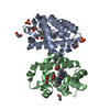
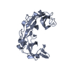
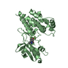
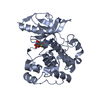
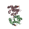
| Structure viewer | Molecule: MolmilJmol/JSmol |
|---|
- Downloads & links
-Download
| PDBx/mmCIF format | 2f3g.cif.gz | 67.5 KB | Display | PDBx/mmCIF format |
|---|---|---|---|---|
| PDB format | pdb2f3g.ent.gz | 50.8 KB | Display | PDB format |
| PDBx/mmJSON format | 2f3g.json.gz | Tree view | PDBx/mmJSON format | |
| Others |  Other downloads Other downloads |
-Validation report
| Arichive directory | https://data.pdbj.org/pub/pdb/validation_reports/f3/2f3gftp://data.pdbj.org/pub/pdb/validation_reports/f3/2f3g | HTTPS FTP |
|---|
-Related structure data
| Related structure data |  1f3zC  1f3gS S: Starting model for refinement C: citing same article ( |
|---|---|
| Similar structure data |


-Links
 PDBj
PDBj- Assembly
| Deposited unit | 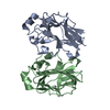
| ||||||||
|---|---|---|---|---|---|---|---|---|---|
| 1 |
| ||||||||
| Unit cell |
| ||||||||
| Noncrystallographic symmetry (NCS) | NCS oper: (Code: given Matrix: (-0.0515, -0.8642, -0.5006), Vector: Details | NONCRYSTALLOGRAPHIC SYMMETRY IS PROBABLY NOT BIOLOGICALLY RELEVANT. | |
-Components
| #1: Protein | Mass: 18141.834 Da / Num. of mol.: 2 / Source method: isolated from a natural source Details: IIAGLC PROTEOLYZED-MISSING 16 N-TERMINAL AMINO ACIDS Source: (natural) References: UniProt: P69783, protein-Npi-phosphohistidine-sugar phosphotransferase #2: Water | ChemComp-HOH / |  Mass: 18.015 Da / Num. of mol.: 59 / Source method: isolated from a natural source / Formula: H2O Mass: 18.015 Da / Num. of mol.: 59 / Source method: isolated from a natural source / Formula: H2O |
|---|
-Experimental details
-Experiment
| Experiment | Method: X-RAY DIFFRACTION / Number of used crystals: 1 |
|---|
- Sample preparation
| Crystal | Density Matthews: 2.17 Å3/Da / Density % sol: 43 % | ||||||||||||||||||||||||||||||
|---|---|---|---|---|---|---|---|---|---|---|---|---|---|---|---|---|---|---|---|---|---|---|---|---|---|---|---|---|---|---|---|
| Crystal grow | pH: 8.5 Details: 30% PEG 4000, 0.2M MGCL2, 0.1M TRIS, PH 8.5 4 DEGREES | ||||||||||||||||||||||||||||||
| Crystal grow | *PLUS Method: vapor diffusion, hanging drop | ||||||||||||||||||||||||||||||
| Components of the solutions | *PLUS
|
-Data collection
| Diffraction | Mean temperature: 293 K |
|---|---|
| Diffraction source | Source: ROTATING ANODE / Type: RIGAKU / Wavelength: 1.5418 |
| Detector | Type: XUONG-HAMLIN MULTIWIRE / Detector: AREA DETECTOR / Date: Jun 1, 1993 |
| Radiation | Monochromator: GRAPHITE(002) / Monochromatic (M) / Laue (L): M / Scattering type: x-ray |
| Radiation wavelength | Wavelength: 1.5418 Å / Relative weight: 1 |
| Reflection | Resolution: 2.13→20 Å / Num. obs: 15616 / % possible obs: 86 % / Observed criterion σ(I): 0 / Redundancy: 2.8 % / Biso Wilson estimate: 21.8 Å2 / Rmerge(I) obs: 0.036 |
| Reflection | *PLUS Num. measured all: 44614 |
- Processing
| Software |
| ||||||||||||||||||||||||||||||||||||||||||||||||||
|---|---|---|---|---|---|---|---|---|---|---|---|---|---|---|---|---|---|---|---|---|---|---|---|---|---|---|---|---|---|---|---|---|---|---|---|---|---|---|---|---|---|---|---|---|---|---|---|---|---|---|---|
| Refinement | Method to determine structure: MOLECULAR REPLACEMENT Starting model: PDB ENTRY 1F3G Resolution: 2.13→20 Å / Isotropic thermal model: TNT BCORREL V2.1 / σ(F): 0 / Stereochemistry target values: TNT PROTGEO
| ||||||||||||||||||||||||||||||||||||||||||||||||||
| Solvent computation | Solvent model: BABINET SCALING / Bsol: 300 Å2 / ksol: 0.8 e/Å3 | ||||||||||||||||||||||||||||||||||||||||||||||||||
| Refinement step | Cycle: LAST / Resolution: 2.13→20 Å
| ||||||||||||||||||||||||||||||||||||||||||||||||||
| Refine LS restraints |
| ||||||||||||||||||||||||||||||||||||||||||||||||||
| Software | *PLUS Name: TNT / Version: 5E / Classification: refinement | ||||||||||||||||||||||||||||||||||||||||||||||||||
| Refinement | *PLUS Rfactor obs: 0.169 | ||||||||||||||||||||||||||||||||||||||||||||||||||
| Solvent computation | *PLUS | ||||||||||||||||||||||||||||||||||||||||||||||||||
| Displacement parameters | *PLUS | ||||||||||||||||||||||||||||||||||||||||||||||||||
| Refine LS restraints | *PLUS
|
