 Movie
Movie Controller


+ Open data

- Basic information
| Entry | Database: PDB / ID: 2qms | ||||||
|---|---|---|---|---|---|---|---|
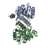
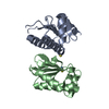
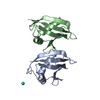
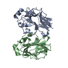
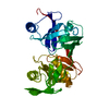
| Title | Crystal structure of a signaling molecule | ||||||
 Components Components | Growth factor receptor-bound protein 7 | ||||||
 Keywords Keywords | SIGNALING PROTEIN / SH2 domain / alpha/beta fold | ||||||
| Function / homology |  Function and homology information Function and homology informationGRB7 events in ERBB2 signaling / RND1 GTPase cycle / RET signaling / cell projection / Tie2 Signaling / phosphatidylinositol binding / Downstream signal transduction / stress granule assembly / Signaling by SCF-KIT / epidermal growth factor receptor signaling pathway ...GRB7 events in ERBB2 signaling / RND1 GTPase cycle / RET signaling / cell projection / Tie2 Signaling / phosphatidylinositol binding / Downstream signal transduction / stress granule assembly / Signaling by SCF-KIT / epidermal growth factor receptor signaling pathway / cytoplasmic stress granule / negative regulation of translation / positive regulation of cell migration / focal adhesion / protein kinase binding / RNA binding / identical protein binding / plasma membrane / cytosol Similarity search - Function | ||||||
| Biological species |  Homo sapiens (human) Homo sapiens (human) | ||||||
| Method |  X-RAY DIFFRACTION / MOLECULAR REPLACEMENT / molecular replacement / Resolution: 2.1 Å X-RAY DIFFRACTION / MOLECULAR REPLACEMENT / molecular replacement / Resolution: 2.1 Å | ||||||
 Authors Authors | Porter, C.J. / Wilce, M.C. / Wilce, J.A. | ||||||
 Citation Citation | Journal: Bmc Struct.Biol. / Year: 2007 Title: Grb7 SH2 domain structure and interactions with a cyclic peptide inhibitor of cancer cell migration and proliferation. Authors: Porter, C.J. / Matthews, J.M. / Mackay, J.P. / Pursglove, S.E. / Schmidberger, J.W. / Leedman, P.J. / Pero, S.C. / Krag, D.N. / Wilce, M.C. / Wilce, J.A. | ||||||
| History |
|
- Structure visualization
| Structure viewer | Molecule: MolmilJmol/JSmol |
|---|
- Downloads & links
-Download
| PDBx/mmCIF format | 2qms.cif.gz | 107.4 KB | Display | PDBx/mmCIF format |
|---|---|---|---|---|
| PDB format | pdb2qms.ent.gz | 84.3 KB | Display | PDB format |
| PDBx/mmJSON format | 2qms.json.gz | Tree view | PDBx/mmJSON format | |
| Others |  Other downloads Other downloads |
-Validation report
| Arichive directory | https://data.pdbj.org/pub/pdb/validation_reports/qm/2qmsftp://data.pdbj.org/pub/pdb/validation_reports/qm/2qms | HTTPS FTP |
|---|
-Related structure data
| Similar structure data |
|---|


-Links
 PDBj
PDBj


- Assembly
| Deposited unit | 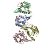
| ||||||||
|---|---|---|---|---|---|---|---|---|---|
| 1 | 
| ||||||||
| 2 | 
| ||||||||
| 3 | 
| ||||||||
| 4 | 
| ||||||||
| Unit cell |
| ||||||||
| Details | The biological unit is a dimer. There are 2 biological units in the asymmetric unit (chains A & B and chains C & D) |
-Components
| #1: Protein | Mass: 13690.711 Da / Num. of mol.: 4 / Fragment: SH2 domain (residues 415-532) Source method: isolated from a genetically manipulated source Source: (gene. exp.) Homo sapiens (human) / Gene: GRB7 / Plasmid: pGex4T2 / Production host:  #2: Chemical | ChemComp-SO4 /   Mass: 96.063 Da / Num. of mol.: 7 / Source method: obtained synthetically / Formula: SO4 Mass: 96.063 Da / Num. of mol.: 7 / Source method: obtained synthetically / Formula: SO4#3: Water | ChemComp-HOH / |  Mass: 18.015 Da / Num. of mol.: 181 / Source method: isolated from a natural source / Formula: H2O Mass: 18.015 Da / Num. of mol.: 181 / Source method: isolated from a natural source / Formula: H2O |
|---|
-Experimental details
-Experiment
| Experiment | Method: X-RAY DIFFRACTION / Number of used crystals: 1 |
|---|
- Sample preparation
| Crystal | Density Matthews: 1.93 Å3/Da / Density % sol: 36.3 % |
|---|---|
| Crystal grow | Temperature: 295 K / Method: vapor diffusion, hanging drop / pH: 6.1 Details: 0.1M sodium citrate pH6.1, 22.5% PEG 4000, 0.2M Ammonium sulphate, 5 % glycerol, VAPOR DIFFUSION, HANGING DROP, temperature 295K |
-Data collection
| Diffraction | Mean temperature: 100 K |
|---|---|
| Diffraction source | Source: ROTATING ANODE / Type: RIGAKU RUH2R / Wavelength: 1.5418 Å |
| Detector | Type: MAR scanner 345 mm plate / Detector: IMAGE PLATE / Date: Jan 1, 2004 / Details: osmic mirrors |
| Radiation | Protocol: SINGLE WAVELENGTH / Scattering type: x-ray |
| Radiation wavelength | Wavelength: 1.5418 Å / Relative weight: 1 |
| Reflection | Resolution: 2.1→44.7 Å / Num. obs: 25401 / % possible obs: 99.1 % / Redundancy: 3.1 % / Biso Wilson estimate: 28.4 Å2 / Rmerge(I) obs: 0.084 / Net I/σ(I): 12.6 |
| Reflection shell | Resolution: 2.1→2.15 Å / Rmerge(I) obs: 0.505 / Mean I/σ(I) obs: 2.2 / Num. unique all: 1269 / % possible all: 99.4 |
-Phasing
| Phasing | Method: molecular replacement | |||||||||
|---|---|---|---|---|---|---|---|---|---|---|
| Phasing MR | Rfactor: 0.502 / Cor.coef. Fo:Fc: 0.544
|
- Processing
| Software |
| ||||||||||||||||||||||||||||||||||||||||||||||||||||||||||||||||||||||||||||||||||||||||||
|---|---|---|---|---|---|---|---|---|---|---|---|---|---|---|---|---|---|---|---|---|---|---|---|---|---|---|---|---|---|---|---|---|---|---|---|---|---|---|---|---|---|---|---|---|---|---|---|---|---|---|---|---|---|---|---|---|---|---|---|---|---|---|---|---|---|---|---|---|---|---|---|---|---|---|---|---|---|---|---|---|---|---|---|---|---|---|---|---|---|---|---|
| Refinement | Method to determine structure: MOLECULAR REPLACEMENT / Resolution: 2.1→30 Å / Cor.coef. Fo:Fc: 0.956 / Cor.coef. Fo:Fc free: 0.928 / SU B: 6.16 / SU ML: 0.166 / Cross valid method: THROUGHOUT / σ(F): 0 / ESU R: 0.291 / ESU R Free: 0.22 / Stereochemistry target values: MAXIMUM LIKELIHOOD / Details: HYDROGENS HAVE BEEN ADDED IN THE RIDING POSITIONS
| ||||||||||||||||||||||||||||||||||||||||||||||||||||||||||||||||||||||||||||||||||||||||||
| Solvent computation | Ion probe radii: 0.8 Å / Shrinkage radii: 0.8 Å / VDW probe radii: 1.4 Å / Solvent model: BABINET MODEL WITH MASK | ||||||||||||||||||||||||||||||||||||||||||||||||||||||||||||||||||||||||||||||||||||||||||
| Displacement parameters | Biso mean: 37.848 Å2
| ||||||||||||||||||||||||||||||||||||||||||||||||||||||||||||||||||||||||||||||||||||||||||
| Refinement step | Cycle: LAST / Resolution: 2.1→30 Å
| ||||||||||||||||||||||||||||||||||||||||||||||||||||||||||||||||||||||||||||||||||||||||||
| Refine LS restraints |
| ||||||||||||||||||||||||||||||||||||||||||||||||||||||||||||||||||||||||||||||||||||||||||
| LS refinement shell | Resolution: 2.1→2.154 Å / Total num. of bins used: 20
|
