Mass: 158.925 Da / Num. of mol.: 1 / Source method: obtained synthetically / Formula: Tb
Compound details
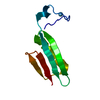
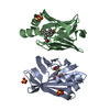
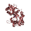
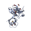
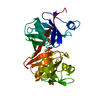
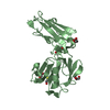
A67, R69, E207, AND A294 ARE MUTATIONS FOR THE PREVENTION OF HOMO-OLIGOMERIZATION. S42, S226, AND ...A67, R69, E207, AND A294 ARE MUTATIONS FOR THE PREVENTION OF HOMO-OLIGOMERIZATION. S42, S226, AND S242 ARE FOR THE FIXATION OF LANTHANIDE-BINDING PEPTIDE TAG(LBT).
Sequence details
THE SEQUENCE CYVDTNNDGAYEGDELHMG OF CHAIN A IS FOR LANTHANIDE-BINDING PEPTIDE SEQUENCE.
Method: DGSA-distance geometry simulated annealing / Software ordinal: 1 Details: In this work, the dimer structure of p62 PB1 was determined by using a rigid-body docking calculation based on the pseudo-contact shift. In the docking calculation, the coordinates of chain ...Details: In this work, the dimer structure of p62 PB1 was determined by using a rigid-body docking calculation based on the pseudo-contact shift. In the docking calculation, the coordinates of chain A were held fixed, whereas the coordinates of chain B were treated as a rigid body and set free to rotate and translate. The differences among the models are relative orientations between chain A and chain B, not each coordinates. In the calculation, monomer structures of DR and KE were docked each other based on the inter-subunit restraints from pseudo-contact shifts. The coordinates of DR and KE were generated from the monomer structure of DR (2kkc), using PyMOL software. For pseudo-contact shift restraints, we need a paramagnetic lanthanide ion fixed in the target protein. To fix a lanthanide ions to DR, we utilized the lanthanide binding peptide tag (LBT).The chain A represents LBT-DR, and chain B does KE. We collected the pseudo-contact shift restrains using LBT-DR/KE complex, while the docking calculations were performed using the coordinates without LBT.
NMR representative
Selection criteria: lowest energy
NMR ensemble
Conformer selection criteria: structures with the lowest energy Conformers calculated total number: 100 / Conformers submitted total number: 10 / Representative conformer: 1
+
About Yorodumi
-
News
-
Feb 9, 2022. New format data for meta-information of EMDB entries
New format data for meta-information of EMDB entries
Version 3 of the EMDB header file is now the official format.
The previous official version 1.9 will be removed from the archive.
In the structure databanks used in Yorodumi, some data are registered as the other names, "COVID-19 virus" and "2019-nCoV". Here are the details of the virus and the list of structure data.
Jan 31, 2019. EMDB accession codes are about to change! (news from PDBe EMDB page)
EMDB accession codes are about to change! (news from PDBe EMDB page)
The allocation of 4 digits for EMDB accession codes will soon come to an end. Whilst these codes will remain in use, new EMDB accession codes will include an additional digit and will expand incrementally as the available range of codes is exhausted. The current 4-digit format prefixed with “EMD-” (i.e. EMD-XXXX) will advance to a 5-digit format (i.e. EMD-XXXXX), and so on. It is currently estimated that the 4-digit codes will be depleted around Spring 2019, at which point the 5-digit format will come into force.
The EM Navigator/Yorodumi systems omit the EMD- prefix.
Related info.:Q: What is EMD? / ID/Accession-code notation in Yorodumi/EM Navigator
Yorodumi is a browser for structure data from EMDB, PDB, SASBDB, etc.
This page is also the successor to EM Navigator detail page, and also detail information page/front-end page for Omokage search.
The word "yorodu" (or yorozu) is an old Japanese word meaning "ten thousand". "mi" (miru) is to see.
Related info.:EMDB / PDB / SASBDB / Comparison of 3 databanks / Yorodumi Search / Aug 31, 2016. New EM Navigator & Yorodumi / Yorodumi Papers / Jmol/JSmol / Function and homology information / Changes in new EM Navigator and Yorodumi
 Movie
Movie Controller


 Open data
 Basic information
 Components
 Keywords
 Function and homology information
 Authors
 Citation
 Structure visualization
 Downloads & links
 Other downloads


 PDBj
PDBj


 Assembly
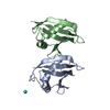

 Mass: 158.925 Da / Num. of mol.: 1 / Source method: obtained synthetically / Formula: Tb
 HSQC
HSQC Sample preparation
 Processing