 Movie
Movie Controller

[English] 日本語
 Yorodumi
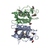
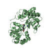
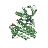
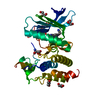
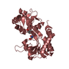
- PDB-3p42: Structure of GfcC (YmcB), protein encoded by the E. coli group 4 ... -
+ Open data

- Basic information
| Entry | Database: PDB / ID: 3p42 | ||||||
|---|---|---|---|---|---|---|---|
| Title | Structure of GfcC (YmcB), protein encoded by the E. coli group 4 capsule operon | ||||||
 Components Components | Predicted protein | ||||||
 Keywords Keywords | UNKNOWN FUNCTION / Beta-Grasp | ||||||
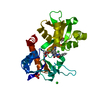
| Function / homology |  Function and homology information Function and homology informationUbiquitin-like (UB roll) - #700 / Single alpha-helices involved in coiled-coils or other helix-helix interfaces - #2280 / Capsule biosynthesis GfcC-like, C-terminal / Capsule biosynthesis GfcC-like, N-terminal / Capsule biosynthesis GfcC C-terminal / Capsule biosynthesis GfcC, N-terminal / Outer membrane lipoprotein wza fold like / Outer membrane lipoprotein wza domain like / Single alpha-helices involved in coiled-coils or other helix-helix interfaces / Helix non-globular ...Ubiquitin-like (UB roll) - #700 / Single alpha-helices involved in coiled-coils or other helix-helix interfaces - #2280 / Capsule biosynthesis GfcC-like, C-terminal / Capsule biosynthesis GfcC-like, N-terminal / Capsule biosynthesis GfcC C-terminal / Capsule biosynthesis GfcC, N-terminal / Outer membrane lipoprotein wza fold like / Outer membrane lipoprotein wza domain like / Single alpha-helices involved in coiled-coils or other helix-helix interfaces / Helix non-globular / Special / Ubiquitin-like (UB roll) / Roll / Alpha Beta Similarity search - Domain/homology | ||||||
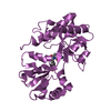
| Biological species |  | ||||||
| Method |  X-RAY DIFFRACTION / SYNCHROTRON / SAD / Resolution: 1.91 Å X-RAY DIFFRACTION / SYNCHROTRON / SAD / Resolution: 1.91 Å | ||||||
 Authors Authors | Saper, M.A. / Sathiyamoorthy, K. | ||||||
 Citation Citation | Journal: Biochemistry / Year: 2011 Title: The Crystal Structure of Escherichia coli Group 4 Capsule Protein GfcC Reveals a Domain Organization Resembling That of Wza. Authors: Sathiyamoorthy, K. / Mills, E. / Franzmann, T.M. / Rosenshine, I. / Saper, M.A. | ||||||
| History |
|
- Structure visualization
| Structure viewer | Molecule: MolmilJmol/JSmol |
|---|
- Downloads & links
-Download
| PDBx/mmCIF format | 3p42.cif.gz | 527.5 KB | Display | PDBx/mmCIF format |
|---|---|---|---|---|
| PDB format | pdb3p42.ent.gz | 443.1 KB | Display | PDB format |
| PDBx/mmJSON format | 3p42.json.gz | Tree view | PDBx/mmJSON format | |
| Others |  Other downloads Other downloads |
-Validation report
| Arichive directory | https://data.pdbj.org/pub/pdb/validation_reports/p4/3p42ftp://data.pdbj.org/pub/pdb/validation_reports/p4/3p42 | HTTPS FTP |
|---|
-Related structure data
| Similar structure data |
|---|


-Links
 PDBj
PDBj- Assembly
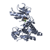
| Deposited unit | 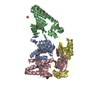
| |||||||||||||||||||||||||||
|---|---|---|---|---|---|---|---|---|---|---|---|---|---|---|---|---|---|---|---|---|---|---|---|---|---|---|---|---|
| 1 | 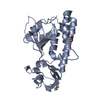
| |||||||||||||||||||||||||||
| 2 | 
| |||||||||||||||||||||||||||
| 3 | 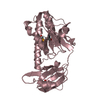
| |||||||||||||||||||||||||||
| 4 | 
| |||||||||||||||||||||||||||
| 5 | 
| |||||||||||||||||||||||||||
| 6 | 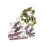
| |||||||||||||||||||||||||||
| Unit cell |
| |||||||||||||||||||||||||||
| Noncrystallographic symmetry (NCS) | NCS domain:
NCS domain segments:
|
-Components
| #1: Protein | Mass: 26266.117 Da / Num. of mol.: 4 / Fragment: UNP residues 22-248 Source method: isolated from a genetically manipulated source Source: (gene. exp.) #2: Chemical |   Mass: 96.063 Da / Num. of mol.: 3 / Source method: obtained synthetically / Formula: SO4 Mass: 96.063 Da / Num. of mol.: 3 / Source method: obtained synthetically / Formula: SO4#3: Water | ChemComp-HOH / |  Mass: 18.015 Da / Num. of mol.: 693 / Source method: isolated from a natural source / Formula: H2O Mass: 18.015 Da / Num. of mol.: 693 / Source method: isolated from a natural source / Formula: H2OHas protein modification | Y | |
|---|
-Experimental details
-Experiment
| Experiment | Method: X-RAY DIFFRACTION / Number of used crystals: 1 |
|---|
- Sample preparation
| Crystal | Density Matthews: 2.26 Å3/Da / Density % sol: 45.56 % |
|---|---|
| Crystal grow | Temperature: 277.15 K / Method: vapor diffusion, sitting drop / pH: 6.5 Details: 1.5 M Ammonium Sulfate, 0.1 M NaCl, 0.1 M Bis-Tris pH 6.5, VAPOR DIFFUSION, SITTING DROP, temperature 277.15K |
-Data collection
| Diffraction | Mean temperature: 100 K |
|---|---|
| Diffraction source | Source: SYNCHROTRON / Site: APS  / Beamline: 21-ID-D / Wavelength: 0.9793 Å / Beamline: 21-ID-D / Wavelength: 0.9793 Å |
| Detector | Type: MARMOSAIC 300 mm CCD / Detector: CCD / Date: Feb 6, 2010 |
| Radiation | Monochromator: Kohzu / Protocol: SINGLE WAVELENGTH / Monochromatic (M) / Laue (L): M / Scattering type: x-ray |
| Radiation wavelength | Wavelength: 0.9793 Å / Relative weight: 1 |
| Reflection | Resolution: 1.77→49 Å / Num. obs: 81677 / Redundancy: 6.8 % / Biso Wilson estimate: 25.01 Å2 |
- Processing
| Software |
| |||||||||||||||||||||||||||||||||||||||||||||||||||||||||||||||||||||||||||||||||||||||||||||||||||||||||||||||||||||||||||||
|---|---|---|---|---|---|---|---|---|---|---|---|---|---|---|---|---|---|---|---|---|---|---|---|---|---|---|---|---|---|---|---|---|---|---|---|---|---|---|---|---|---|---|---|---|---|---|---|---|---|---|---|---|---|---|---|---|---|---|---|---|---|---|---|---|---|---|---|---|---|---|---|---|---|---|---|---|---|---|---|---|---|---|---|---|---|---|---|---|---|---|---|---|---|---|---|---|---|---|---|---|---|---|---|---|---|---|---|---|---|---|---|---|---|---|---|---|---|---|---|---|---|---|---|---|---|---|
| Refinement | Method to determine structure: SAD / Resolution: 1.91→34.494 Å / Occupancy max: 1 / Occupancy min: 0.25 / FOM work R set: 0.8582 / SU ML: 0.25 / σ(F): 0 / Phase error: 21.93 / Stereochemistry target values: ML Details: Refinement was with anomalous data, F+ and F- were refined as separate reflections.
| |||||||||||||||||||||||||||||||||||||||||||||||||||||||||||||||||||||||||||||||||||||||||||||||||||||||||||||||||||||||||||||
| Solvent computation | Shrinkage radii: 0.53 Å / VDW probe radii: 0.7 Å / Solvent model: FLAT BULK SOLVENT MODEL / Bsol: 51.964 Å2 / ksol: 0.433 e/Å3 | |||||||||||||||||||||||||||||||||||||||||||||||||||||||||||||||||||||||||||||||||||||||||||||||||||||||||||||||||||||||||||||
| Displacement parameters | Biso max: 242.89 Å2 / Biso mean: 40.8623 Å2 / Biso min: 11.59 Å2
| |||||||||||||||||||||||||||||||||||||||||||||||||||||||||||||||||||||||||||||||||||||||||||||||||||||||||||||||||||||||||||||
| Refinement step | Cycle: LAST / Resolution: 1.91→34.494 Å
| |||||||||||||||||||||||||||||||||||||||||||||||||||||||||||||||||||||||||||||||||||||||||||||||||||||||||||||||||||||||||||||
| Refine LS restraints |
| |||||||||||||||||||||||||||||||||||||||||||||||||||||||||||||||||||||||||||||||||||||||||||||||||||||||||||||||||||||||||||||
| Refine LS restraints NCS |
| |||||||||||||||||||||||||||||||||||||||||||||||||||||||||||||||||||||||||||||||||||||||||||||||||||||||||||||||||||||||||||||
| LS refinement shell | Refine-ID: X-RAY DIFFRACTION / Total num. of bins used: 10
| |||||||||||||||||||||||||||||||||||||||||||||||||||||||||||||||||||||||||||||||||||||||||||||||||||||||||||||||||||||||||||||
| Refinement TLS params. | Method: refined / Refine-ID: X-RAY DIFFRACTION
| |||||||||||||||||||||||||||||||||||||||||||||||||||||||||||||||||||||||||||||||||||||||||||||||||||||||||||||||||||||||||||||
| Refinement TLS group |
|
