 Movie
Movie Controller

[English] 日本語
 Yorodumi
- PDB-1k46: Crystal Structure of the Type III Secretory Domain of Yersinia Yo... -
+ Open data

- Basic information
| Entry | Database: PDB / ID: 1k46 | ||||||
|---|---|---|---|---|---|---|---|
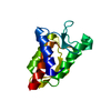
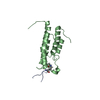
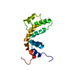
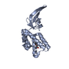
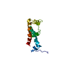
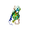
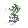
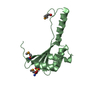
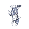
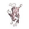
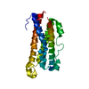
| Title | Crystal Structure of the Type III Secretory Domain of Yersinia YopH Reveals a Domain-Swapped Dimer | ||||||
 Components Components | PROTEIN-TYROSINE PHOSPHATASE YOPH | ||||||
 Keywords Keywords | HYDROLASE / domain-swap / phosphopeptide-binding domain / type III secretion domain | ||||||
| Function / homology |  Function and homology information Function and homology informationprotein-tyrosine-phosphatase / protein tyrosine phosphatase activity / extracellular region Similarity search - Function | ||||||
| Biological species |  Yersinia pseudotuberculosis (bacteria) Yersinia pseudotuberculosis (bacteria) | ||||||
| Method |  X-RAY DIFFRACTION / MIR / Resolution: 2.2 Å X-RAY DIFFRACTION / MIR / Resolution: 2.2 Å | ||||||
 Authors Authors | Smith, C.L. / Khandelwal, P. / Keliikuli, K. / Zuiderweg, E.R.P. / Saper, M.A. | ||||||
 Citation Citation | Journal: Mol.Microbiol. / Year: 2001 Title: Structure of the type III secretion and substrate-binding domain of Yersinia YopH phosphatase. Authors: Smith, C.L. / Khandelwal, P. / Keliikuli, K. / Zuiderweg, E.R. / Saper, M.A. #1: Journal: J.Biol.Chem. / Year: 2001Title: Identification of Residues in the N-terminal Domain of the Yersinia Tyrosine Phosphatase that are Critical for Substrate Recognition Authors: Montagna, L.G. / Ivanov, M.I. / Bliska, J.B. #2: Journal: Mol.Microbiol. / Year: 1998Title: Identification of an Amino-terminal Substrate-binding Domain in the Yersinia Tyrosine Phosphatase that is Required for Efficient Recognition of Focal Adhesion Targets Authors: Black, D.S. / Montagna, L.G. / Zitsmann, S. / Bliska, J.B. | ||||||
| History |
| ||||||
| Remark 300 | BIOMOLECULE: 1 THIS ENTRY CONTAINS THE CRYSTALLOGRAPHIC ASYMMETRIC UNIT WHICH CONSISTS OF 1 CHAIN(S) ...BIOMOLECULE: 1 THIS ENTRY CONTAINS THE CRYSTALLOGRAPHIC ASYMMETRIC UNIT WHICH CONSISTS OF 1 CHAIN(S). THIS IS LIKELY THE BIOLOGICALLY-RELEVANT STRUCTURE. THE RELEVANCE OF THE DOMAIN-SWAPPED DIMER OBSERVED IN THE CRYSTAL STRUCTURE IS DESCRIBED IN THE JRNL REFERENCE. ALSO SEE REMARK 900. THE OTHER MOLECULE OF THE DOMAIN-SWAPPED DIMER IS GENERATED BY APPLYING CRYSTALLOGRAPHIC OPERATOR 3 IN REMARK 290 FOLLOWED BY A TRANSLATION OF 47.90 IN X. |
- Structure visualization
| Structure viewer | Molecule: MolmilJmol/JSmol |
|---|
- Downloads & links
-Download
| PDBx/mmCIF format | 1k46.cif.gz | 38.2 KB | Display | PDBx/mmCIF format |
|---|---|---|---|---|
| PDB format | pdb1k46.ent.gz | 26.9 KB | Display | PDB format |
| PDBx/mmJSON format | 1k46.json.gz | Tree view | PDBx/mmJSON format | |
| Others |  Other downloads Other downloads |
-Validation report
| Arichive directory | https://data.pdbj.org/pub/pdb/validation_reports/k4/1k46ftp://data.pdbj.org/pub/pdb/validation_reports/k4/1k46 | HTTPS FTP |
|---|
-Related structure data
| Related structure data | |
|---|---|
| Similar structure data |
-Links
 PDBj
PDBj


- Assembly
| Deposited unit | 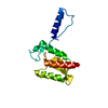
| ||||||||
|---|---|---|---|---|---|---|---|---|---|
| 1 |
| ||||||||
| 2 | 
| ||||||||
| Unit cell |
| ||||||||
| Details | YopH(1-129) can exist as a monomer or dimer in solution. Crystal structure is a domain-swapped dimer consisting of two molecules related by a crystallographic two fold axis: -x+1,y,-z+1/2. The physiologically-relevant form is likely the monomer. |
-Components
| #1: Protein | Mass: 14882.599 Da / Num. of mol.: 1 / Fragment: amino-terminal domain (residues 1-129) Source method: isolated from a genetically manipulated source Source: (gene. exp.) Yersinia pseudotuberculosis (bacteria) / Gene: yopH / Plasmid: pT7-7 / Species (production host): Escherichia coli / Production host: |
|---|---|
| #2: Water | ChemComp-HOH /  Mass: 18.015 Da / Num. of mol.: 110 / Source method: isolated from a natural source / Formula: H2O Mass: 18.015 Da / Num. of mol.: 110 / Source method: isolated from a natural source / Formula: H2O |
-Experimental details
-Experiment
| Experiment | Method: X-RAY DIFFRACTION / Number of used crystals: 1 |
|---|
- Sample preparation
| Crystal | Density Matthews: 2.37 Å3/Da / Density % sol: 48.09 % | |||||||||||||||||||||||||||||||||||||||||||||||||||||||||||||||
|---|---|---|---|---|---|---|---|---|---|---|---|---|---|---|---|---|---|---|---|---|---|---|---|---|---|---|---|---|---|---|---|---|---|---|---|---|---|---|---|---|---|---|---|---|---|---|---|---|---|---|---|---|---|---|---|---|---|---|---|---|---|---|---|---|
| Crystal grow | Temperature: 295 K / Method: vapor diffusion, hanging drop / pH: 8 Details: PEG 8000, sodium chlolride, Tris , VAPOR DIFFUSION, HANGING DROP, temperature 295K | |||||||||||||||||||||||||||||||||||||||||||||||||||||||||||||||
| Crystal grow | *PLUS Temperature: 22 ℃ / pH: 7.2 / Method: vapor diffusion | |||||||||||||||||||||||||||||||||||||||||||||||||||||||||||||||
| Components of the solutions | *PLUS
|
-Data collection
| Diffraction | Mean temperature: 120 K |
|---|---|
| Diffraction source | Source: ROTATING ANODE / Type: RIGAKU RUH3R / Wavelength: 1.5418 Å |
| Detector | Type: RIGAKU RAXIS IV / Detector: IMAGE PLATE / Date: Aug 11, 1998 / Details: mirrors |
| Radiation | Monochromator: Yale double focusing mirrors / Protocol: SINGLE WAVELENGTH / Monochromatic (M) / Laue (L): M / Scattering type: x-ray |
| Radiation wavelength | Wavelength: 1.5418 Å / Relative weight: 1 |
| Reflection | Resolution: 2.2→22 Å / Num. all: 7503 / Num. obs: 7503 / % possible obs: 99.8 % / Observed criterion σ(F): 0 / Observed criterion σ(I): 0 / Redundancy: 6.2 % / Biso Wilson estimate: 23.3 Å2 / Rmerge(I) obs: 0.05 / Rsym value: 0.05 / Net I/σ(I): 20.8 |
| Reflection shell | Resolution: 2.2→2.28 Å / Redundancy: 6.1 % / Rmerge(I) obs: 0.136 / Mean I/σ(I) obs: 16.2 / Num. unique all: 720 / % possible all: 99.9 |
| Reflection | *PLUS Num. measured all: 68285 / Rmerge(I) obs: 0.05 |
| Reflection shell | *PLUS % possible obs: 99.9 % / Num. unique obs: 720 |
- Processing
| Software |
| |||||||||||||||||||||||||
|---|---|---|---|---|---|---|---|---|---|---|---|---|---|---|---|---|---|---|---|---|---|---|---|---|---|---|
| Refinement | Method to determine structure: MIR Starting model: de novo Resolution: 2.2→22 Å / Isotropic thermal model: isotropic / Cross valid method: R-free / σ(F): 0 / σ(I): 0 / Stereochemistry target values: Engh & Huber
| |||||||||||||||||||||||||
| Displacement parameters | Biso mean: 30.8 Å2 | |||||||||||||||||||||||||
| Refinement step | Cycle: LAST / Resolution: 2.2→22 Å
| |||||||||||||||||||||||||
| Refine LS restraints |
| |||||||||||||||||||||||||
| LS refinement shell | Resolution: 2.2→2.25 Å
| |||||||||||||||||||||||||
| Software | *PLUS Name: CNS / Version: 0.9 / Classification: refinement | |||||||||||||||||||||||||
| Refinement | *PLUS Lowest resolution: 22 Å / σ(F): 0 / Rfactor obs: 0.224 / Rfactor Rfree: 0.258 | |||||||||||||||||||||||||
| Solvent computation | *PLUS | |||||||||||||||||||||||||
| Displacement parameters | *PLUS Biso mean: 30.8 Å2 | |||||||||||||||||||||||||
| LS refinement shell | *PLUS Rfactor Rwork: 0.222 / Rfactor obs: 0.222 |
