 Movie
Movie Controller

[English] 日本語
 Yorodumi
- PDB-1huf: CRYSTAL STRUCTURE OF THE N-TERMINAL DOMAIN OF THE TYROSINE PHOSPH... -
+ Open data

- Basic information
| Entry | Database: PDB / ID: 1huf | ||||||
|---|---|---|---|---|---|---|---|
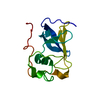
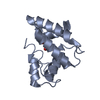
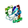
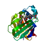
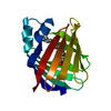
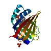
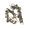
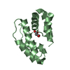
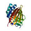
| Title | CRYSTAL STRUCTURE OF THE N-TERMINAL DOMAIN OF THE TYROSINE PHOSPHATASE YOPH FROM YERSINIA PESTIS. | ||||||
 Components Components | TYROSINE PHOSPHATASE YOPH | ||||||
 Keywords Keywords | HYDROLASE / helical bundle / beta hairpin | ||||||
| Function / homology |  Function and homology information Function and homology informationsymbiont-mediated suppression of host reactive oxygen species generation / symbiont-mediated disruption of host focal adhesion / protein-tyrosine-phosphatase / protein tyrosine phosphatase activity / extracellular region Similarity search - Function | ||||||
| Biological species |   Yersinia pestis (bacteria) Yersinia pestis (bacteria) | ||||||
| Method |  X-RAY DIFFRACTION / MIR / Resolution: 2 Å X-RAY DIFFRACTION / MIR / Resolution: 2 Å | ||||||
 Authors Authors | Evdokimov, A.G. / Waugh, D.S. | ||||||
 Citation Citation | Journal: Acta Crystallogr.,Sect.D / Year: 2001 Title: Structure of the N-terminal domain of Yersinia pestis YopH at 2.0 A resolution. Authors: Evdokimov, A.G. / Tropea, J.E. / Routzahn, K.M. / Copeland, T.D. / Waugh, D.S. | ||||||
| History |
|
- Structure visualization
| Structure viewer | Molecule: MolmilJmol/JSmol |
|---|
- Downloads & links
-Download
| PDBx/mmCIF format | 1huf.cif.gz | 36.9 KB | Display | PDBx/mmCIF format |
|---|---|---|---|---|
| PDB format | pdb1huf.ent.gz | 25.7 KB | Display | PDB format |
| PDBx/mmJSON format | 1huf.json.gz | Tree view | PDBx/mmJSON format | |
| Others |  Other downloads Other downloads |
-Validation report
| Arichive directory | https://data.pdbj.org/pub/pdb/validation_reports/hu/1hufftp://data.pdbj.org/pub/pdb/validation_reports/hu/1huf | HTTPS FTP |
|---|
-Related structure data
| Related structure data | |
|---|---|
| Similar structure data |


-Links
 PDBj
PDBj


- Assembly
| Deposited unit | 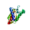
| ||||||||||||
|---|---|---|---|---|---|---|---|---|---|---|---|---|---|
| 1 |
| ||||||||||||
| Unit cell |
| ||||||||||||
| Components on special symmetry positions |
|
-Components
| #1: Protein | Mass: 15080.780 Da / Num. of mol.: 1 / Fragment: N-TERMINAL DOMAIN Source method: isolated from a genetically manipulated source Source: (gene. exp.) Yersinia pestis (bacteria) / Gene: YOPH / Plasmid: PKM904 / Production host: |
|---|---|
| #2: Water | ChemComp-HOH /  Mass: 18.015 Da / Num. of mol.: 94 / Source method: isolated from a natural source / Formula: H2O Mass: 18.015 Da / Num. of mol.: 94 / Source method: isolated from a natural source / Formula: H2O |
-Experimental details
-Experiment
| Experiment | Method: X-RAY DIFFRACTION / Number of used crystals: 1 |
|---|
- Sample preparation
| Crystal | Density Matthews: 2.46 Å3/Da / Density % sol: 49.71 % | ||||||||||||||||||
|---|---|---|---|---|---|---|---|---|---|---|---|---|---|---|---|---|---|---|---|
| Crystal grow | Temperature: 293 K / Method: vapor diffusion, hanging drop / pH: 9 Details: PEG4000, bicine, pH 9, VAPOR DIFFUSION, HANGING DROP, temperature 293K | ||||||||||||||||||
| Crystal grow | *PLUS pH: 8 / Method: vapor diffusion | ||||||||||||||||||
| Components of the solutions | *PLUS
|
-Data collection
| Diffraction | Mean temperature: 100 K |
|---|---|
| Diffraction source | Source: ROTATING ANODE / Type: RIGAKU / Wavelength: 1.54178 |
| Detector | Type: MARRESEARCH / Detector: IMAGE PLATE / Date: Nov 5, 2000 / Details: Osmic mirrors |
| Radiation | Monochromator: graphite / Protocol: SINGLE WAVELENGTH / Monochromatic (M) / Laue (L): M / Scattering type: x-ray |
| Radiation wavelength | Wavelength: 1.54178 Å / Relative weight: 1 |
| Reflection | Resolution: 2→100 Å / Num. all: 9659 / Num. obs: 8860 / % possible obs: 83.3 % / Observed criterion σ(F): 2 / Observed criterion σ(I): 2 / Redundancy: 4.01 % / Biso Wilson estimate: 21.2 Å2 / Rmerge(I) obs: 0.072 / Rsym value: 0.045 / Net I/σ(I): 15.3 |
| Reflection shell | Resolution: 2.01→2.08 Å / Redundancy: 3.6 % / Rmerge(I) obs: 0.34 / Mean I/σ(I) obs: 3.6 / Num. unique all: 711 / % possible all: 75.3 |
| Reflection | *PLUS Lowest resolution: 100 Å |
| Reflection shell | *PLUS % possible obs: 55 % |
- Processing
| Software |
| |||||||||||||||||||||||||
|---|---|---|---|---|---|---|---|---|---|---|---|---|---|---|---|---|---|---|---|---|---|---|---|---|---|---|
| Refinement | Method to determine structure: MIR / Resolution: 2→100 Å / Isotropic thermal model: isotropic one B factor per atom / Cross valid method: THROUGHOUT / σ(F): 2 / σ(I): 2 / Stereochemistry target values: Engh & Huber Details: One simulated annealing run (CNS) followed by conjugate-gradient least squares procedure. Data cutoff of 2 sigma is 8046 reflections.
| |||||||||||||||||||||||||
| Solvent computation | Solvent model: Babinet (SHELXL) / Bsol: 250 Å2 / ksol: 0.83 e/Å3 | |||||||||||||||||||||||||
| Displacement parameters | Biso mean: 31 Å2 | |||||||||||||||||||||||||
| Refinement step | Cycle: LAST / Resolution: 2→100 Å
| |||||||||||||||||||||||||
| Refine LS restraints |
| |||||||||||||||||||||||||
| Software | *PLUS Name: 'CNS, SHELXL-97' / Classification: refinement | |||||||||||||||||||||||||
| Refinement | *PLUS Highest resolution: 2 Å / Lowest resolution: 100 Å / σ(F): 2 / % reflection Rfree: 8 % / Rfactor Rfree: 0.26 | |||||||||||||||||||||||||
| Solvent computation | *PLUS | |||||||||||||||||||||||||
| Displacement parameters | *PLUS Biso mean: 31 Å2 | |||||||||||||||||||||||||
| Refine LS restraints | *PLUS
|
