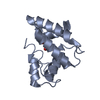
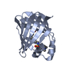
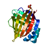
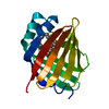
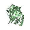
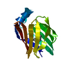
- PDB-2nnq: Crystal structure of human adipocyte fatty acid binding protein i... -
+
Open data
ID or keywords:
Loading...
-
Basic information
Entry
Database: PDB / ID: 2nnq
Title
Crystal structure of human adipocyte fatty acid binding protein in complex with ((2'-(5-ethyl-3,4-diphenyl-1H-pyrazol-1-yl)-3-biphenylyl)oxy)acetic acid
Components
Fatty acid-binding protein, adipocyte
Keywords
LIPID TRANSPORT / TRANSPORT / LIPID-BINDING
Function / homology
Function and homology information
white fat cell proliferation / hormone receptor binding / long-chain fatty acid transmembrane transporter activity / long-chain fatty acid binding / cellular response to lithium ion / Triglyceride catabolism / white fat cell differentiation / long-chain fatty acid transport / fatty acid transport / lipid droplet ...white fat cell proliferation / hormone receptor binding / long-chain fatty acid transmembrane transporter activity / long-chain fatty acid binding / cellular response to lithium ion / Triglyceride catabolism / white fat cell differentiation / long-chain fatty acid transport / fatty acid transport / lipid droplet / brown fat cell differentiation / fatty acid binding / cholesterol homeostasis / response to bacterium / Transcriptional regulation of white adipocyte differentiation / cellular response to tumor necrosis factor / positive regulation of inflammatory response / positive regulation of cold-induced thermogenesis / MLL4 and MLL3 complexes regulate expression of PPARG target genes in adipogenesis and hepatic steatosis / negative regulation of DNA-templated transcription / extracellular exosome / nucleus / cytoplasm / cytosol Similarity search - Function
In the structure databanks used in Yorodumi, some data are registered as the other names, "COVID-19 virus" and "2019-nCoV". Here are the details of the virus and the list of structure data.
Jan 31, 2019. EMDB accession codes are about to change! (news from PDBe EMDB page)
EMDB accession codes are about to change! (news from PDBe EMDB page)
The allocation of 4 digits for EMDB accession codes will soon come to an end. Whilst these codes will remain in use, new EMDB accession codes will include an additional digit and will expand incrementally as the available range of codes is exhausted. The current 4-digit format prefixed with “EMD-” (i.e. EMD-XXXX) will advance to a 5-digit format (i.e. EMD-XXXXX), and so on. It is currently estimated that the 4-digit codes will be depleted around Spring 2019, at which point the 5-digit format will come into force.
The EM Navigator/Yorodumi systems omit the EMD- prefix.
Related info.:Q: What is EMD? / ID/Accession-code notation in Yorodumi/EM Navigator
Yorodumi is a browser for structure data from EMDB, PDB, SASBDB, etc.
This page is also the successor to EM Navigator detail page, and also detail information page/front-end page for Omokage search.
The word "yorodu" (or yorozu) is an old Japanese word meaning "ten thousand". "mi" (miru) is to see.
Related info.:EMDB / PDB / SASBDB / Comparison of 3 databanks / Yorodumi Search / Aug 31, 2016. New EM Navigator & Yorodumi / Yorodumi Papers / Jmol/JSmol / Function and homology information / Changes in new EM Navigator and Yorodumi
 Movie
Movie Controller

 Yorodumi
 Open data
 Basic information
 Components
 Keywords
 Function and homology information
 Homo sapiens (human)
 X-RAY DIFFRACTION /
  Authors
 Citation
 Structure visualization
 Downloads & links
 Other downloads
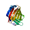

 PDBj
PDBj

 Assembly
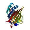


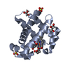
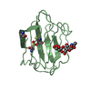
 Mass: 96.063 Da / Num. of mol.: 2 / Source method: obtained synthetically / Formula: SO4
 Mass: 474.550 Da / Num. of mol.: 1 / Source method: obtained synthetically / Formula: C31H26N2O3
 Mass: 18.015 Da / Num. of mol.: 152 / Source method: isolated from a natural source / Formula: H2O
 Sample preparation
 Processing