 Movie
Movie Controller

[English] 日本語
 Yorodumi
- PDB-5edc: human FABP4 in complex with 6-Chloro-4-phenyl-2-piperidin-1-yl-qu... -
+ Open data

- Basic information
| Entry | Database: PDB / ID: 5edc | ||||||
|---|---|---|---|---|---|---|---|
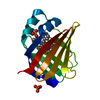
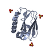
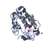
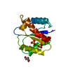
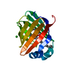
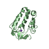
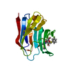
| Title | human FABP4 in complex with 6-Chloro-4-phenyl-2-piperidin-1-yl-quinoline-3-carboxylic acid at 1.29A | ||||||
 Components Components | Fatty acid-binding protein, adipocyte | ||||||
 Keywords Keywords | LIPID BINDING PROTEIN / FATTY ACID BINDING PROTEIN / CYTOPLASM / LIPID-BINDING / TRANSPORT / PROTEIN BINDING | ||||||
| Function / homology |  Function and homology information Function and homology informationwhite fat cell proliferation / hormone receptor binding / long-chain fatty acid transmembrane transporter activity / cellular response to lithium ion / long-chain fatty acid binding / Triglyceride catabolism / white fat cell differentiation / long-chain fatty acid transport / fatty acid transport / lipid droplet ...white fat cell proliferation / hormone receptor binding / long-chain fatty acid transmembrane transporter activity / cellular response to lithium ion / long-chain fatty acid binding / Triglyceride catabolism / white fat cell differentiation / long-chain fatty acid transport / fatty acid transport / lipid droplet / response to bacterium / brown fat cell differentiation / cholesterol homeostasis / fatty acid binding / Transcriptional regulation of white adipocyte differentiation / cellular response to tumor necrosis factor / positive regulation of inflammatory response / positive regulation of cold-induced thermogenesis / MLL4 and MLL3 complexes regulate expression of PPARG target genes in adipogenesis and hepatic steatosis / negative regulation of DNA-templated transcription / extracellular exosome / nucleus / cytosol / cytoplasm Similarity search - Function | ||||||
| Biological species |  Homo sapiens (human) Homo sapiens (human) | ||||||
| Method |  X-RAY DIFFRACTION / SYNCHROTRON / Resolution: 1.29 Å X-RAY DIFFRACTION / SYNCHROTRON / Resolution: 1.29 Å | ||||||
 Authors Authors | Rudolph, M.G. / Ehler, A. | ||||||
 Citation Citation | Journal: J.Med.Chem. / Year: 2016 Title: A Real-World Perspective on Molecular Design. Authors: Kuhn, B. / Guba, W. / Hert, J. / Banner, D. / Bissantz, C. / Ceccarelli, S. / Haap, W. / Korner, M. / Kuglstatter, A. / Lerner, C. / Mattei, P. / Neidhart, W. / Pinard, E. / Rudolph, M.G. / ...Authors: Kuhn, B. / Guba, W. / Hert, J. / Banner, D. / Bissantz, C. / Ceccarelli, S. / Haap, W. / Korner, M. / Kuglstatter, A. / Lerner, C. / Mattei, P. / Neidhart, W. / Pinard, E. / Rudolph, M.G. / Schulz-Gasch, T. / Woltering, T. / Stahl, M. | ||||||
| History |
|
- Structure visualization
| Structure viewer | Molecule: MolmilJmol/JSmol |
|---|
- Downloads & links
-Download
| PDBx/mmCIF format | 5edc.cif.gz | 77.5 KB | Display | PDBx/mmCIF format |
|---|---|---|---|---|
| PDB format | pdb5edc.ent.gz | 56.5 KB | Display | PDB format |
| PDBx/mmJSON format | 5edc.json.gz | Tree view | PDBx/mmJSON format | |
| Others |  Other downloads Other downloads |
-Validation report
| Arichive directory | https://data.pdbj.org/pub/pdb/validation_reports/ed/5edcftp://data.pdbj.org/pub/pdb/validation_reports/ed/5edc | HTTPS FTP |
|---|
-Related structure data
| Related structure data |  5edbC 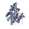 5edeC 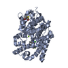 5edgC 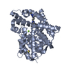 5edhC  5ediC  5ezxC  5ezzC  5f00C 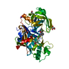 5f01C  5f02C  5f03C  5i2rC C: citing same article ( |
|---|---|
| Similar structure data |


-Links
 PDBj
PDBj

- Assembly
| Deposited unit | 
| ||||||||
|---|---|---|---|---|---|---|---|---|---|
| 1 |
| ||||||||
| Unit cell |
|
-Components
| #1: Protein | Mass: 14810.978 Da / Num. of mol.: 1 / Fragment: SOLUBLE FORM, RESIDUES 3-132 Source method: isolated from a genetically manipulated source Source: (gene. exp.) Homo sapiens (human) / Gene: FABP4 / Production host:  | ||
|---|---|---|---|
| #2: Chemical | ChemComp-5M7 /   Mass: 366.841 Da / Num. of mol.: 1 / Source method: obtained synthetically / Formula: C21H19ClN2O2 Mass: 366.841 Da / Num. of mol.: 1 / Source method: obtained synthetically / Formula: C21H19ClN2O2 | ||
| #3: Chemical |   Mass: 96.063 Da / Num. of mol.: 3 / Source method: obtained synthetically / Formula: SO4 Mass: 96.063 Da / Num. of mol.: 3 / Source method: obtained synthetically / Formula: SO4#4: Water | ChemComp-HOH / |  Mass: 18.015 Da / Num. of mol.: 125 / Source method: isolated from a natural source / Formula: H2O Mass: 18.015 Da / Num. of mol.: 125 / Source method: isolated from a natural source / Formula: H2O |
-Experimental details
-Experiment
| Experiment | Method: X-RAY DIFFRACTION / Number of used crystals: 1 |
|---|
- Sample preparation
| Crystal | Density Matthews: 2.18 Å3/Da / Density % sol: 43.69 % |
|---|---|
| Crystal grow | Temperature: 295 K / Method: vapor diffusion, sitting drop Details: 0.1 M HEPES/NaOH pH 7.5, 2.0-2.2 M ammonium sulfate |
-Data collection
| Diffraction | Mean temperature: 100 K | ||||||||||||||||||||||||||||||||||||||||||||||||||||||||||||||||||||||||||||||||||||||||||||||||||||||||||||||||||||||||||||||||||||||||||||||||||||||||||||||||||||||||||||||||||||||||||||||||||||||||||||||||||
|---|---|---|---|---|---|---|---|---|---|---|---|---|---|---|---|---|---|---|---|---|---|---|---|---|---|---|---|---|---|---|---|---|---|---|---|---|---|---|---|---|---|---|---|---|---|---|---|---|---|---|---|---|---|---|---|---|---|---|---|---|---|---|---|---|---|---|---|---|---|---|---|---|---|---|---|---|---|---|---|---|---|---|---|---|---|---|---|---|---|---|---|---|---|---|---|---|---|---|---|---|---|---|---|---|---|---|---|---|---|---|---|---|---|---|---|---|---|---|---|---|---|---|---|---|---|---|---|---|---|---|---|---|---|---|---|---|---|---|---|---|---|---|---|---|---|---|---|---|---|---|---|---|---|---|---|---|---|---|---|---|---|---|---|---|---|---|---|---|---|---|---|---|---|---|---|---|---|---|---|---|---|---|---|---|---|---|---|---|---|---|---|---|---|---|---|---|---|---|---|---|---|---|---|---|---|---|---|---|---|---|---|
| Diffraction source | Source: SYNCHROTRON / Site: SLS  / Beamline: X10SA / Wavelength: 1 Å / Beamline: X10SA / Wavelength: 1 Å | ||||||||||||||||||||||||||||||||||||||||||||||||||||||||||||||||||||||||||||||||||||||||||||||||||||||||||||||||||||||||||||||||||||||||||||||||||||||||||||||||||||||||||||||||||||||||||||||||||||||||||||||||||
| Detector | Type: PSI PILATUS 6M / Detector: PIXEL / Date: Nov 12, 2010 | ||||||||||||||||||||||||||||||||||||||||||||||||||||||||||||||||||||||||||||||||||||||||||||||||||||||||||||||||||||||||||||||||||||||||||||||||||||||||||||||||||||||||||||||||||||||||||||||||||||||||||||||||||
| Radiation | Protocol: SINGLE WAVELENGTH / Monochromatic (M) / Laue (L): M / Scattering type: x-ray | ||||||||||||||||||||||||||||||||||||||||||||||||||||||||||||||||||||||||||||||||||||||||||||||||||||||||||||||||||||||||||||||||||||||||||||||||||||||||||||||||||||||||||||||||||||||||||||||||||||||||||||||||||
| Radiation wavelength | Wavelength: 1 Å / Relative weight: 1 | ||||||||||||||||||||||||||||||||||||||||||||||||||||||||||||||||||||||||||||||||||||||||||||||||||||||||||||||||||||||||||||||||||||||||||||||||||||||||||||||||||||||||||||||||||||||||||||||||||||||||||||||||||
| Reflection | Resolution: 1.24→43.51 Å / Num. obs: 37129 / % possible obs: 98.6 % / Observed criterion σ(I): -3 / Redundancy: 6.08 % / Biso Wilson estimate: 22.666 Å2 / CC1/2: 0.999 / Rmerge(I) obs: 0.055 / Rrim(I) all: 0.06 / Χ2: 1.037 / Net I/σ(I): 12.86 / Num. measured all: 228777 | ||||||||||||||||||||||||||||||||||||||||||||||||||||||||||||||||||||||||||||||||||||||||||||||||||||||||||||||||||||||||||||||||||||||||||||||||||||||||||||||||||||||||||||||||||||||||||||||||||||||||||||||||||
| Reflection shell | Diffraction-ID: 1 / Rejects: _
|
- Processing
| Software |
| ||||||||||||||||||||||||
|---|---|---|---|---|---|---|---|---|---|---|---|---|---|---|---|---|---|---|---|---|---|---|---|---|---|
| Refinement | Resolution: 1.29→43.51 Å / Cor.coef. Fo:Fc: 0.976 / Cor.coef. Fo:Fc free: 0.97 / SU B: 2.399 / SU ML: 0.043 / Cross valid method: THROUGHOUT / σ(F): 0 / ESU R: 0.054 / ESU R Free: 0.052 / Stereochemistry target values: MAXIMUM LIKELIHOOD Details: HYDROGENS HAVE BEEN USED IF PRESENT IN THE INPUT U VALUES : REFINED INDIVIDUALLY
| ||||||||||||||||||||||||
| Solvent computation | Ion probe radii: 0.8 Å / Shrinkage radii: 0.8 Å / VDW probe radii: 1.2 Å / Solvent model: BABINET MODEL WITH MASK | ||||||||||||||||||||||||
| Displacement parameters | Biso max: 76.05 Å2 / Biso mean: 19.711 Å2 / Biso min: 12.6 Å2
| ||||||||||||||||||||||||
| Refinement step | Cycle: final / Resolution: 1.29→43.51 Å
| ||||||||||||||||||||||||
| LS refinement shell | Resolution: 1.29→1.323 Å / Total num. of bins used: 20
|
