 Movie
Movie Controller

[English] 日本語
 Yorodumi
- PDB-5edh: CRYSTAL STRUCTURE OF HUMAN PHOSPHODIESTERASE 10 IN COMPLEX WITH n... -
+ Open data

- Basic information
| Entry | Database: PDB / ID: 5edh | ||||||
|---|---|---|---|---|---|---|---|
| Title | CRYSTAL STRUCTURE OF HUMAN PHOSPHODIESTERASE 10 IN COMPLEX WITH n4c(C)n1c(nc(n1)CCc2nc(nn2C)N3CCCC3)c(c4)CC, micromolar IC50=0.0037753 | ||||||
 Components Components | cAMP and cAMP-inhibited cGMP 3',5'-cyclic phosphodiesterase 10A | ||||||
 Keywords Keywords | HYDROLASE / PHOSPHODIESTERASE / PDE10 | ||||||
| Function / homology |  Function and homology information Function and homology information3',5'-cGMP-stimulated cyclic-nucleotide phosphodiesterase activity / 3',5'-cyclic-nucleotide phosphodiesterase / negative regulation of receptor guanylyl cyclase signaling pathway / cGMP catabolic process / cAMP catabolic process / cGMP effects / 3',5'-cyclic-nucleotide phosphodiesterase activity / cGMP binding / 3',5'-cyclic-GMP phosphodiesterase activity / 3',5'-cyclic-AMP phosphodiesterase activity ...3',5'-cGMP-stimulated cyclic-nucleotide phosphodiesterase activity / 3',5'-cyclic-nucleotide phosphodiesterase / negative regulation of receptor guanylyl cyclase signaling pathway / cGMP catabolic process / cAMP catabolic process / cGMP effects / 3',5'-cyclic-nucleotide phosphodiesterase activity / cGMP binding / 3',5'-cyclic-GMP phosphodiesterase activity / 3',5'-cyclic-AMP phosphodiesterase activity / regulation of adenylate cyclase-activating G protein-coupled receptor signaling pathway / cAMP binding / negative regulation of cAMP/PKA signal transduction / G alpha (s) signalling events / glutamatergic synapse / signal transduction / metal ion binding / cytosol Similarity search - Function | ||||||
| Biological species |  Homo sapiens (human) Homo sapiens (human) | ||||||
| Method |  X-RAY DIFFRACTION / SYNCHROTRON / MOLECULAR REPLACEMENT / Resolution: 2.03 Å X-RAY DIFFRACTION / SYNCHROTRON / MOLECULAR REPLACEMENT / Resolution: 2.03 Å | ||||||
 Authors Authors | Joseph, C. / Rudolph, M.G. | ||||||
 Citation Citation | Journal: J.Med.Chem. / Year: 2016 Title: A Real-World Perspective on Molecular Design. Authors: Kuhn, B. / Guba, W. / Hert, J. / Banner, D. / Bissantz, C. / Ceccarelli, S. / Haap, W. / Korner, M. / Kuglstatter, A. / Lerner, C. / Mattei, P. / Neidhart, W. / Pinard, E. / Rudolph, M.G. / ...Authors: Kuhn, B. / Guba, W. / Hert, J. / Banner, D. / Bissantz, C. / Ceccarelli, S. / Haap, W. / Korner, M. / Kuglstatter, A. / Lerner, C. / Mattei, P. / Neidhart, W. / Pinard, E. / Rudolph, M.G. / Schulz-Gasch, T. / Woltering, T. / Stahl, M. | ||||||
| History |
|
- Structure visualization
| Structure viewer | Molecule: MolmilJmol/JSmol |
|---|
- Downloads & links
-Download
| PDBx/mmCIF format | 5edh.cif.gz | 274 KB | Display | PDBx/mmCIF format |
|---|---|---|---|---|
| PDB format | pdb5edh.ent.gz | 221.1 KB | Display | PDB format |
| PDBx/mmJSON format | 5edh.json.gz | Tree view | PDBx/mmJSON format | |
| Others |  Other downloads Other downloads |
-Validation report
| Arichive directory | https://data.pdbj.org/pub/pdb/validation_reports/ed/5edhftp://data.pdbj.org/pub/pdb/validation_reports/ed/5edh | HTTPS FTP |
|---|
-Related structure data
| Related structure data |  5edbC  5edcC  5edeC  5edgC  5ediC  5ezxC  5ezzC  5f00C  5f01C  5f02C  5f03C  5i2rC C: citing same article ( |
|---|---|
| Similar structure data |

-Links
 PDBj
PDBj

- Assembly
| Deposited unit | 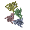
| ||||||||
|---|---|---|---|---|---|---|---|---|---|
| 1 | 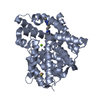
| ||||||||
| 2 | 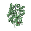
| ||||||||
| 3 | 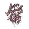
| ||||||||
| 4 | 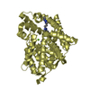
| ||||||||
| Unit cell |
|
-Components
| #1: Protein | Mass: 36515.152 Da / Num. of mol.: 4 Source method: isolated from a genetically manipulated source Source: (gene. exp.) Homo sapiens (human) / Gene: PDE10A / Production host:  References: UniProt: Q9Y233, 3',5'-cyclic-nucleotide phosphodiesterase, 3',5'-cyclic-GMP phosphodiesterase #2: Chemical | ChemComp-ZN /   Mass: 65.409 Da / Num. of mol.: 4 / Source method: obtained synthetically / Formula: Zn Mass: 65.409 Da / Num. of mol.: 4 / Source method: obtained synthetically / Formula: Zn#3: Chemical | ChemComp-MG /   Mass: 24.305 Da / Num. of mol.: 4 / Source method: obtained synthetically / Formula: Mg Mass: 24.305 Da / Num. of mol.: 4 / Source method: obtained synthetically / Formula: Mg#4: Chemical | ChemComp-5MF /   Mass: 340.426 Da / Num. of mol.: 4 / Source method: obtained synthetically / Formula: C17H24N8 Mass: 340.426 Da / Num. of mol.: 4 / Source method: obtained synthetically / Formula: C17H24N8#5: Water | ChemComp-HOH / |  Mass: 18.015 Da / Num. of mol.: 526 / Source method: isolated from a natural source / Formula: H2O Mass: 18.015 Da / Num. of mol.: 526 / Source method: isolated from a natural source / Formula: H2OHas protein modification | Y | |
|---|
-Experimental details
-Experiment
| Experiment | Method: X-RAY DIFFRACTION / Number of used crystals: 1 |
|---|
- Sample preparation
| Crystal | Density Matthews: 2.83 Å3/Da / Density % sol: 56.59 % |
|---|---|
| Crystal grow | Temperature: 295 K / Method: vapor diffusion, sitting drop / Details: 0.1 M HEPES-NaOH pH 7.5, 30% PEG550MME, 50mM MgCl2 |
-Data collection
| Diffraction | Mean temperature: 100 K |
|---|---|
| Diffraction source | Source: SYNCHROTRON / Site: SLS  / Beamline: X10SA / Wavelength: 0.99995 Å / Beamline: X10SA / Wavelength: 0.99995 Å |
| Detector | Type: DECTRIS PILATUS 6M / Detector: PIXEL / Date: May 17, 2013 |
| Radiation | Protocol: SINGLE WAVELENGTH / Monochromatic (M) / Laue (L): M / Scattering type: x-ray |
| Radiation wavelength | Wavelength: 0.99995 Å / Relative weight: 1 |
| Reflection | Resolution: 2.03→43.68 Å / Num. obs: 103725 / % possible obs: 99.8 % / Observed criterion σ(I): -3 / Redundancy: 5.17 % / Biso Wilson estimate: 44.61 Å2 / CC1/2: 0.999 / Rmerge(I) obs: 0.069 / Rrim(I) all: 0.084 / Rsym value: 0.069 / Χ2: 0.988 / Net I/σ(I): 14.8 / Num. measured all: 548572 |
| Reflection shell | Resolution: 2.03→2.13 Å / Redundancy: 5.05 % / Rmerge(I) obs: 0.742 / Mean I/σ(I) obs: 1.28 / Num. measured obs: 6324 / Num. possible: 1183 / Num. unique obs: 1173 / CC1/2: 1 / Rrim(I) all: 0.018 / Rsym value: 0.742 / Rejects: 0 / % possible all: 99.6 |
- Processing
| Software |
| ||||||||||||||||||||||||
|---|---|---|---|---|---|---|---|---|---|---|---|---|---|---|---|---|---|---|---|---|---|---|---|---|---|
| Refinement | Method to determine structure: MOLECULAR REPLACEMENT / Resolution: 2.03→43.68 Å / Cor.coef. Fo:Fc: 0.966 / Cor.coef. Fo:Fc free: 0.95 / SU B: 4.759 / SU ML: 0.123 / Cross valid method: THROUGHOUT / σ(F): 0 / ESU R: 0.178 / ESU R Free: 0.16 / Stereochemistry target values: MAXIMUM LIKELIHOOD Details: HYDROGENS HAVE BEEN USED IF PRESENT IN THE INPUT U VALUES : REFINED INDIVIDUALLY
| ||||||||||||||||||||||||
| Solvent computation | Ion probe radii: 0.8 Å / Shrinkage radii: 0.8 Å / VDW probe radii: 1.2 Å / Solvent model: MASK | ||||||||||||||||||||||||
| Displacement parameters | Biso max: 80 Å2 / Biso mean: 40.871 Å2 / Biso min: 18.84 Å2
| ||||||||||||||||||||||||
| Refinement step | Cycle: final / Resolution: 2.03→43.68 Å
| ||||||||||||||||||||||||
| LS refinement shell | Resolution: 2.03→2.083 Å / Total num. of bins used: 20
|
