 Movie
Movie Controller

[English] 日本語
 Yorodumi
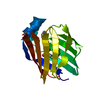
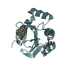
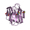
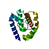
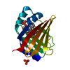
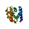
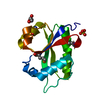
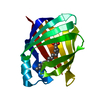
- PDB-3stm: Structure of human LFABP in complex with one molecule of palmitic acid -
+ Open data

- Basic information
| Entry | Database: PDB / ID: 3stm | ||||||
|---|---|---|---|---|---|---|---|
| Title | Structure of human LFABP in complex with one molecule of palmitic acid | ||||||
 Components Components | Fatty acid-binding protein, liver | ||||||
 Keywords Keywords | LIPID BINDING PROTEIN / LFABP / Palmitic acid / Fatty acid binding | ||||||
| Function / homology |  Function and homology information Function and homology informationcellular detoxification / Heme degradation / Triglyceride catabolism / antioxidant activity / peroxisomal matrix / fatty acid transport / Regulation of lipid metabolism by PPARalpha / fatty acid binding / PPARA activates gene expression / Cytoprotection by HMOX1 ...cellular detoxification / Heme degradation / Triglyceride catabolism / antioxidant activity / peroxisomal matrix / fatty acid transport / Regulation of lipid metabolism by PPARalpha / fatty acid binding / PPARA activates gene expression / Cytoprotection by HMOX1 / cellular response to hydrogen peroxide / cellular response to hypoxia / chromatin binding / extracellular exosome / nucleoplasm / nucleus / cytosol Similarity search - Function | ||||||
| Biological species |  Homo sapiens (human) Homo sapiens (human) | ||||||
| Method |  X-RAY DIFFRACTION / MOLECULAR REPLACEMENT / molecular replacement / Resolution: 2.216 Å X-RAY DIFFRACTION / MOLECULAR REPLACEMENT / molecular replacement / Resolution: 2.216 Å | ||||||
 Authors Authors | Sharma, A. / Sharma, A. | ||||||
 Citation Citation | Journal: J.Biol.Chem. / Year: 2011 Title: Fatty acid induced remodeling within the Human liver fatty acid binding protein Authors: Sharma, A. / Sharma, A. | ||||||
| History |
|
- Structure visualization
| Structure viewer | Molecule: MolmilJmol/JSmol |
|---|
- Downloads & links
-Download
| PDBx/mmCIF format | 3stm.cif.gz | 39.3 KB | Display | PDBx/mmCIF format |
|---|---|---|---|---|
| PDB format | pdb3stm.ent.gz | 26.1 KB | Display | PDB format |
| PDBx/mmJSON format | 3stm.json.gz | Tree view | PDBx/mmJSON format | |
| Others |  Other downloads Other downloads |
-Validation report
| Arichive directory | https://data.pdbj.org/pub/pdb/validation_reports/st/3stmftp://data.pdbj.org/pub/pdb/validation_reports/st/3stm | HTTPS FTP |
|---|
-Related structure data


-Links
 PDBj
PDBj


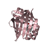
- Assembly
| Deposited unit | 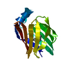
| ||||||||
|---|---|---|---|---|---|---|---|---|---|
| 1 |
| ||||||||
| Unit cell |
|
-Components
| #1: Protein | Mass: 14599.728 Da / Num. of mol.: 1 Source method: isolated from a genetically manipulated source Source: (gene. exp.) Homo sapiens (human) / Gene: FABP1, FABPL / Production host:  |
|---|---|
| #2: Chemical | ChemComp-PLM /   Mass: 256.424 Da / Num. of mol.: 1 / Source method: obtained synthetically / Formula: C16H32O2 Mass: 256.424 Da / Num. of mol.: 1 / Source method: obtained synthetically / Formula: C16H32O2 |
| #3: Water | ChemComp-HOH /  Mass: 18.015 Da / Num. of mol.: 66 / Source method: isolated from a natural source / Formula: H2O Mass: 18.015 Da / Num. of mol.: 66 / Source method: isolated from a natural source / Formula: H2O |
-Experimental details
-Experiment
| Experiment | Method: X-RAY DIFFRACTION / Number of used crystals: 1 |
|---|
- Sample preparation
| Crystal | Density Matthews: 2.21 Å3/Da / Density % sol: 44.37 % |
|---|---|
| Crystal grow | Temperature: 293 K / Method: vapor diffusion, hanging drop / pH: 8 Details: 0.15 M potassium bromide and 30% polyethylene glycol monomethyl ether (PEG MME) 2000, pH 8.0, VAPOR DIFFUSION, HANGING DROP, temperature 293K |
-Data collection
| Diffraction | Mean temperature: 100 K |
|---|---|
| Diffraction source | Source: ROTATING ANODE / Type: RIGAKU MICROMAX-007 |
| Detector | Type: MAR scanner 345 mm plate / Detector: IMAGE PLATE / Date: May 7, 2010 |
| Radiation | Protocol: SINGLE WAVELENGTH / Monochromatic (M) / Laue (L): M / Scattering type: x-ray |
| Radiation wavelength | Relative weight: 1 |
| Reflection | Resolution: 2.216→28.011 Å / Num. obs: 6817 / % possible obs: 98.2 % / Observed criterion σ(F): 2 / Observed criterion σ(I): -3 |
| Reflection shell | Resolution: 2.21→2.29 Å / Redundancy: 5 % / Rmerge(I) obs: 0.45 / % possible all: 86.5 |
-Phasing
| Phasing | Method: molecular replacement |
|---|
- Processing
| Software |
| ||||||||||||||||||||||||||||||||
|---|---|---|---|---|---|---|---|---|---|---|---|---|---|---|---|---|---|---|---|---|---|---|---|---|---|---|---|---|---|---|---|---|---|
| Refinement | Method to determine structure: MOLECULAR REPLACEMENT / Resolution: 2.216→19.176 Å / Occupancy max: 1 / Occupancy min: 0.48 / FOM work R set: 0.7322 / SU ML: 0.33 / σ(F): 1.35 / Phase error: 31.81 / Stereochemistry target values: ML
| ||||||||||||||||||||||||||||||||
| Solvent computation | Shrinkage radii: 0.83 Å / VDW probe radii: 1.1 Å / Solvent model: FLAT BULK SOLVENT MODEL / Bsol: 35.744 Å2 / ksol: 0.278 e/Å3 | ||||||||||||||||||||||||||||||||
| Displacement parameters | Biso max: 69.31 Å2 / Biso mean: 40.3881 Å2 / Biso min: 23.85 Å2
| ||||||||||||||||||||||||||||||||
| Refinement step | Cycle: LAST / Resolution: 2.216→19.176 Å
| ||||||||||||||||||||||||||||||||
| Refine LS restraints |
| ||||||||||||||||||||||||||||||||
| LS refinement shell | Refine-ID: X-RAY DIFFRACTION / Total num. of bins used: 2 / % reflection obs: 99 %
|
