 Movie
Movie Controller

[English] 日本語
 Yorodumi
- PDB-2dpm: DPNM DNA ADENINE METHYLTRANSFERASE FROM STREPTOCCOCUS PNEUMONIAE ... -
+ Open data

- Basic information
| Entry | Database: PDB / ID: 2dpm | ||||||
|---|---|---|---|---|---|---|---|
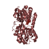
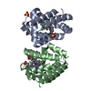
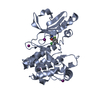
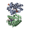
| Title | DPNM DNA ADENINE METHYLTRANSFERASE FROM STREPTOCCOCUS PNEUMONIAE COMPLEXED WITH S-ADENOSYLMETHIONINE | ||||||
 Components Components | PROTEIN (ADENINE-SPECIFIC METHYLTRANSFERASE DPNII 1) | ||||||
 Keywords Keywords | TRANSFERASE / DNA ADENINE METHYLTRANSFERASE / METHYLTRANSFERASE / METHYLASE / DNA METHYLTRANSFERASE GROUP ALPHA / S-ADENOSYLMETHIONINE / DNA N6-ADENINE METHYLATION / DPNII RESTRICTION-MODIFICATION SYSTEM / GATC METHYLATION / ROSSMANN FOLD | ||||||
| Function / homology |  Function and homology information Function and homology informationsite-specific DNA-methyltransferase (adenine-specific) / site-specific DNA-methyltransferase (adenine-specific) activity / S-adenosyl-L-methionine binding / DNA restriction-modification system / mismatch repair / methylation / sequence-specific DNA binding Similarity search - Function | ||||||
| Biological species |   Streptococcus pneumoniae (bacteria) Streptococcus pneumoniae (bacteria) | ||||||
| Method |  X-RAY DIFFRACTION / SYNCHROTRON / MAD / Resolution: 1.8 Å X-RAY DIFFRACTION / SYNCHROTRON / MAD / Resolution: 1.8 Å | ||||||
 Authors Authors | Tran, P.H. / Korszun, Z.R. / Cerritelli, S. / Springhorn, S.S. / Lacks, S.A. | ||||||
 Citation Citation | Journal: Structure / Year: 1998 Title: Crystal structure of the DpnM DNA adenine methyltransferase from the DpnII restriction system of streptococcus pneumoniae bound to S-adenosylmethionine. Authors: Tran, P.H. / Korszun, Z.R. / Cerritelli, S. / Springhorn, S.S. / Lacks, S.A. #1: Journal: J.Mol.Biol. / Year: 1989Title: Crystallization of the DpnM Methylase from the DpnII Restriction System of Streptococcus Pneumoniae Authors: Cerritelli, S. / White, S.W. / Lacks, S.A. #2: Journal: J.Mol.Biol. / Year: 1987Title: Proteins Encoded by the DpnII Restriction Gene Cassette. Two Methylases and an Endonuclease Authors: de la Campa, A.G. / Kale, P. / Springhorn, S.S. / Lacks, S.A. #3: Journal: Cell(Cambridge,Mass.) / Year: 1986Title: Genetic Basis of the Complementary DpnI and DpnII Restriction Systems of S.pneumoniae: An Intercellular Cassette Mechanism Authors: Lacks, S.A. / Mannarelli, B.M. / Springhorn, S.S. / Greenberg, B. #4: Journal: Proc.Natl.Acad.Sci.USA / Year: 1985Title: Nucleotide Sequence of the Dpn II DNA Methylase Gene of Streptococcus Pneumoniae and its Relationship to the dam Gene of Escherichia Coli Authors: Mannarelli, B.M. / Balganesh, T.S. / Greenberg, B. / Springhorn, S.S. / Lacks, S.A. | ||||||
| History |
|
- Structure visualization
| Structure viewer | Molecule: MolmilJmol/JSmol |
|---|
- Downloads & links
-Download
| PDBx/mmCIF format | 2dpm.cif.gz | 70.4 KB | Display | PDBx/mmCIF format |
|---|---|---|---|---|
| PDB format | pdb2dpm.ent.gz | 51.9 KB | Display | PDB format |
| PDBx/mmJSON format | 2dpm.json.gz | Tree view | PDBx/mmJSON format | |
| Others |  Other downloads Other downloads |
-Validation report
| Arichive directory | https://data.pdbj.org/pub/pdb/validation_reports/dp/2dpmftp://data.pdbj.org/pub/pdb/validation_reports/dp/2dpm | HTTPS FTP |
|---|
-Related structure data
| Similar structure data |
|---|


-Links
 PDBj
PDBj


- Assembly
| Deposited unit | 
| ||||||||
|---|---|---|---|---|---|---|---|---|---|
| 1 |
| ||||||||
| Unit cell |
|
-Components
| #1: Protein | Mass: 32948.371 Da / Num. of mol.: 1 Source method: isolated from a genetically manipulated source Details: S-ADENOSYLMETHIONINE / Source: (gene. exp.) Streptococcus pneumoniae (bacteria) / Strain: HB264 / Gene: DPNM / Plasmid: PLS211 / Gene (production host): DPNM / Production host: References: UniProt: P04043, site-specific DNA-methyltransferase (adenine-specific) |
|---|---|
| #2: Chemical | ChemComp-HG /   Mass: 200.590 Da / Num. of mol.: 1 / Source method: obtained synthetically / Formula: Hg Mass: 200.590 Da / Num. of mol.: 1 / Source method: obtained synthetically / Formula: Hg |
| #3: Chemical | ChemComp-SAM /   Mass: 398.437 Da / Num. of mol.: 1 / Source method: obtained synthetically / Formula: C15H22N6O5S Mass: 398.437 Da / Num. of mol.: 1 / Source method: obtained synthetically / Formula: C15H22N6O5S |
| #4: Water | ChemComp-HOH /  Mass: 18.015 Da / Num. of mol.: 178 / Source method: isolated from a natural source / Formula: H2O Mass: 18.015 Da / Num. of mol.: 178 / Source method: isolated from a natural source / Formula: H2O |
-Experimental details
-Experiment
| Experiment | Method: X-RAY DIFFRACTION / Number of used crystals: 2 |
|---|
- Sample preparation
| Crystal | Density Matthews: 2.49 Å3/Da / Density % sol: 48 % / Description: MAD DATA WERE TREATED AS MIRAS | ||||||||||||||||||||||||||||||||||||||||||||||||||||||
|---|---|---|---|---|---|---|---|---|---|---|---|---|---|---|---|---|---|---|---|---|---|---|---|---|---|---|---|---|---|---|---|---|---|---|---|---|---|---|---|---|---|---|---|---|---|---|---|---|---|---|---|---|---|---|---|
| Crystal grow | Method: vapor diffusion, hanging drop / pH: 7 Details: FOR VAPOR DIFFUSION AT 4 C, HANGING DROPS CONTAINED 0.45 NM 0.9 NMOLS OF SAM, 100 MM HEPES BUFFER, PH 7.0, 250 MM NACL, 5% PEG 3350 IN 6 MICROLITERS. RESEVOIRS CONTAINED 1.0 ML OF 50.0 MM ...Details: FOR VAPOR DIFFUSION AT 4 C, HANGING DROPS CONTAINED 0.45 NM 0.9 NMOLS OF SAM, 100 MM HEPES BUFFER, PH 7.0, 250 MM NACL, 5% PEG 3350 IN 6 MICROLITERS. RESEVOIRS CONTAINED 1.0 ML OF 50.0 MM HEPES, 125 MM NACL, 2.5% PEG 3350., vapor diffusion - hanging drop | ||||||||||||||||||||||||||||||||||||||||||||||||||||||
| Crystal | *PLUS | ||||||||||||||||||||||||||||||||||||||||||||||||||||||
| Crystal grow | *PLUS Temperature: 4 ℃ | ||||||||||||||||||||||||||||||||||||||||||||||||||||||
| Components of the solutions | *PLUS
|
-Data collection
| Diffraction | Mean temperature: 100 K | |||||||||||||||
|---|---|---|---|---|---|---|---|---|---|---|---|---|---|---|---|---|
| Diffraction source | Source: SYNCHROTRON / Site: NSLS  / Beamline: X12C / Wavelength: 1.008, 1.011, 0.997, 0.968 / Beamline: X12C / Wavelength: 1.008, 1.011, 0.997, 0.968 | |||||||||||||||
| Detector | Type: MARRESEARCH / Detector: IMAGE PLATE / Date: May 15, 1996 / Details: MIRRORS | |||||||||||||||
| Radiation | Protocol: MAD / Monochromatic (M) / Laue (L): M / Scattering type: x-ray | |||||||||||||||
| Radiation wavelength |
| |||||||||||||||
| Reflection | Resolution: 1.8→20 Å / Num. obs: 30274 / % possible obs: 99.9 % / Redundancy: 7 % / Rsym value: 0.05 / Net I/σ(I): 33.4 | |||||||||||||||
| Reflection shell | Resolution: 1.8→1.84 Å / Redundancy: 7 % / Mean I/σ(I) obs: 8.7 / Rsym value: 0.22 / % possible all: 100 | |||||||||||||||
| Reflection | *PLUS Num. measured all: 207177 / Rmerge(I) obs: 0.05 | |||||||||||||||
| Reflection shell | *PLUS % possible obs: 100 % / Num. unique obs: 1979 / Num. measured obs: 13527 / Rmerge(I) obs: 0.22 |
- Processing
| Software |
| ||||||||||||||||||||||||||||||||||||||||||||||||||||||||||||
|---|---|---|---|---|---|---|---|---|---|---|---|---|---|---|---|---|---|---|---|---|---|---|---|---|---|---|---|---|---|---|---|---|---|---|---|---|---|---|---|---|---|---|---|---|---|---|---|---|---|---|---|---|---|---|---|---|---|---|---|---|---|
| Refinement | Method to determine structure: MAD / Resolution: 1.8→20 Å / Data cutoff high absF: 10000000 / Data cutoff low absF: 0 / Cross valid method: THROUGHOUT / σ(F): 0
| ||||||||||||||||||||||||||||||||||||||||||||||||||||||||||||
| Refinement step | Cycle: LAST / Resolution: 1.8→20 Å
| ||||||||||||||||||||||||||||||||||||||||||||||||||||||||||||
| Refine LS restraints |
| ||||||||||||||||||||||||||||||||||||||||||||||||||||||||||||
| LS refinement shell | Resolution: 1.8→1.88 Å / Total num. of bins used: 8
| ||||||||||||||||||||||||||||||||||||||||||||||||||||||||||||
| Xplor file | Serial no: 1 / Param file: PARHCSDX.PRO | ||||||||||||||||||||||||||||||||||||||||||||||||||||||||||||
| Software | *PLUS Name: X-PLOR / Version: 3.8 / Classification: refinement | ||||||||||||||||||||||||||||||||||||||||||||||||||||||||||||
| Refinement | *PLUS Highest resolution: 1.8 Å / Lowest resolution: 20 Å / σ(F): 0 / % reflection Rfree: 10 % | ||||||||||||||||||||||||||||||||||||||||||||||||||||||||||||
| Solvent computation | *PLUS | ||||||||||||||||||||||||||||||||||||||||||||||||||||||||||||
| Displacement parameters | *PLUS | ||||||||||||||||||||||||||||||||||||||||||||||||||||||||||||
| Refine LS restraints | *PLUS
| ||||||||||||||||||||||||||||||||||||||||||||||||||||||||||||
| LS refinement shell | *PLUS Rfactor Rfree: 0.336 / % reflection Rfree: 10.2 % / Rfactor Rwork: 0.304 |
