 Movie
Movie Controller

[English] 日本語
 Yorodumi
- PDB-2eax: Crystal structure of human PGRP-IBETAC in complex with glycosamyl... -
+ Open data

- Basic information
| Entry | Database: PDB / ID: 2eax | |||||||||
|---|---|---|---|---|---|---|---|---|---|---|
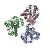
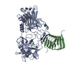
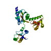
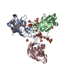
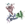
| Title | Crystal structure of human PGRP-IBETAC in complex with glycosamyl muramyl pentapeptide | |||||||||
 Components Components |
| |||||||||
 Keywords Keywords | PEPTIDOGLYCAN-BINDING PROTEIN / ALPHA/BETA | |||||||||
| Function / homology |  Function and homology information Function and homology informationpeptidoglycan immune receptor activity / N-acetylmuramoyl-L-alanine amidase activity / peptidoglycan binding / Antimicrobial peptides / detection of bacterium / peptidoglycan catabolic process / antimicrobial humoral immune response mediated by antimicrobial peptide / killing of cells of another organism / defense response to Gram-positive bacterium / defense response to bacterium ...peptidoglycan immune receptor activity / N-acetylmuramoyl-L-alanine amidase activity / peptidoglycan binding / Antimicrobial peptides / detection of bacterium / peptidoglycan catabolic process / antimicrobial humoral immune response mediated by antimicrobial peptide / killing of cells of another organism / defense response to Gram-positive bacterium / defense response to bacterium / immune response / protein heterodimerization activity / innate immune response / protein-containing complex / extracellular region / zinc ion binding / membrane Similarity search - Function | |||||||||
| Biological species |  Homo sapiens (human) Homo sapiens (human) | |||||||||
| Method |  X-RAY DIFFRACTION / MOLECULAR REPLACEMENT / Resolution: 2.1 Å X-RAY DIFFRACTION / MOLECULAR REPLACEMENT / Resolution: 2.1 Å | |||||||||
 Authors Authors | Cho, S. | |||||||||
 Citation Citation | Journal: Proc.Natl.Acad.Sci.Usa / Year: 2007 Title: Structural insights into the bactericidal mechanism of human peptidoglycan recognition proteins Authors: Cho, S. / Wang, Q. / Swaminathan, C.P. / Hesek, D. / Lee, M. / Boons, G.J. / Mobashery, S. / Mariuzza, R.A. | |||||||||
| History |
|
- Structure visualization
| Structure viewer | Molecule: MolmilJmol/JSmol |
|---|
- Downloads & links
-Download
| PDBx/mmCIF format | 2eax.cif.gz | 115 KB | Display | PDBx/mmCIF format |
|---|---|---|---|---|
| PDB format | pdb2eax.ent.gz | 88 KB | Display | PDB format |
| PDBx/mmJSON format | 2eax.json.gz | Tree view | PDBx/mmJSON format | |
| Others |  Other downloads Other downloads |
-Validation report
| Summary document | 2eax_validation.pdf.gz | 880 KB | Display | wwPDB validaton report |
|---|---|---|---|---|
| Full document | 2eax_full_validation.pdf.gz | 892.7 KB | Display | |
| Data in XML | 2eax_validation.xml.gz | 24.8 KB | Display | |
| Data in CIF | 2eax_validation.cif.gz | 35.7 KB | Display | |
| Arichive directory | https://data.pdbj.org/pub/pdb/validation_reports/ea/2eaxftp://data.pdbj.org/pub/pdb/validation_reports/ea/2eax | HTTPS FTP |
-Related structure data
| Related structure data |  2eavSC S: Starting model for refinement C: citing same article ( |
|---|---|
| Similar structure data |


-Links
 PDBj
PDBj

- Assembly
| Deposited unit | 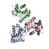
| ||||||||
|---|---|---|---|---|---|---|---|---|---|
| 1 |
| ||||||||
| Unit cell |
|
-Components
| #1: Protein | Mass: 17922.479 Da / Num. of mol.: 3 / Fragment: peptidoglycan-binding domain Source method: isolated from a genetically manipulated source Source: (gene. exp.) Homo sapiens (human) / Gene: PGRPIB / Plasmid: PT7-7 / Species (production host): Escherichia coli / Production host:  #2: Protein/peptide | | Mass: 489.542 Da / Num. of mol.: 1 / Source method: obtained synthetically / Details: The sequence of the peptide is naturally in human. / Source: (synth.) Homo sapiens (human)#3: Polysaccharide | 2-acetamido-2-deoxy-beta-D-glucopyranose-(1-4)-methyl 2-acetamido-3-O-[(1R)-1-carboxyethyl]-2-deoxy- ...2-acetamido-2-deoxy-beta-D-glucopyranose-(1-4)-methyl 2-acetamido-3-O-[(1R)-1-carboxyethyl]-2-deoxy-beta-D-glucopyranoside | #4: Water | ChemComp-HOH / |  Mass: 18.015 Da / Num. of mol.: 320 / Source method: isolated from a natural source / Formula: H2O Mass: 18.015 Da / Num. of mol.: 320 / Source method: isolated from a natural source / Formula: H2OCompound details | THE ENTIRE CHAIN L (RESIDUES 500-506) IS GLYCOSAMYL MURAMYL PENTAPEPTIDE. THE SEQUENCE IS NAG AMV ...THE ENTIRE CHAIN L (RESIDUES 500-506) IS GLYCOSAMYL | Has protein modification | Y | |
|---|
-Experimental details
-Experiment
| Experiment | Method: X-RAY DIFFRACTION / Number of used crystals: 1 |
|---|
- Sample preparation
| Crystal | Density Matthews: 2.31 Å3/Da / Density % sol: 46.74 % |
|---|---|
| Crystal grow | Temperature: 298 K / Method: vapor diffusion, hanging drop / pH: 7 Details: 15% (v/v) Tacsimate, 0.1M Hepes pH 7.0, 2% (w/v) PEG 3350, VAPOR DIFFUSION, HANGING DROP, temperature 298.0K |
-Data collection
| Diffraction | Mean temperature: 100 K |
|---|---|
| Diffraction source | Source: ROTATING ANODE / Type: RIGAKU RU200 / Wavelength: 1.5418 Å |
| Detector | Type: RIGAKU RAXIS IV / Detector: IMAGE PLATE / Date: Aug 15, 2006 / Details: mirror |
| Radiation | Monochromator: SAGITALLY FOCUSED SI(111) / Protocol: SINGLE WAVELENGTH / Monochromatic (M) / Laue (L): M / Scattering type: x-ray |
| Radiation wavelength | Wavelength: 1.5418 Å / Relative weight: 1 |
| Reflection | Resolution: 2.1→50 Å / Num. all: 27838 / Num. obs: 27509 / % possible obs: 97.9 % / Observed criterion σ(F): 2 / Observed criterion σ(I): 2 / Redundancy: 5.2 % / Rmerge(I) obs: 0.064 |
| Reflection shell | Resolution: 2.1→2.16 Å / Rmerge(I) obs: 0.273 / Num. unique all: 1328 / % possible all: 96.72 |
- Processing
| Software |
| |||||||||||||||||||||||||
|---|---|---|---|---|---|---|---|---|---|---|---|---|---|---|---|---|---|---|---|---|---|---|---|---|---|---|
| Refinement | Method to determine structure: MOLECULAR REPLACEMENT Starting model: PDB ENTRY 2EAV Resolution: 2.1→30 Å / σ(F): 2 / σ(I): 4 / Stereochemistry target values: Engh & Huber
| |||||||||||||||||||||||||
| Refinement step | Cycle: LAST / Resolution: 2.1→30 Å
| |||||||||||||||||||||||||
| LS refinement shell | Resolution: 2.1→2.16 Å
|
