 Movie
Movie Controller

[English] 日本語
 Yorodumi
- PDB-6u6f: The crystal structure of anti-apoptotic Mcl-1 protein in complex ... -
+ Open data

- Basic information
| Entry | Database: PDB / ID: 6u6f | ||||||||||||
|---|---|---|---|---|---|---|---|---|---|---|---|---|---|
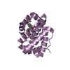
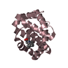
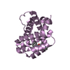
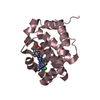
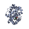
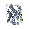
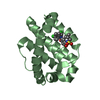
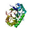
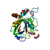
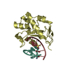
| Title | The crystal structure of anti-apoptotic Mcl-1 protein in complex with 2, 5-substituted benzoic acid inhibitor 21 | ||||||||||||
 Components Components | Induced myeloid leukemia cell differentiation protein Mcl-1 | ||||||||||||
 Keywords Keywords | APOPTOSIS/INHIBITOR / Apoptosis / Mcl1 / Bfl1/A1 / protein-protein interaction / BH3 mimetics / cancer / APOPTOSIS-INHIBITOR complex | ||||||||||||
| Function / homology |  Function and homology information Function and homology informationpositive regulation of oxidative stress-induced neuron intrinsic apoptotic signaling pathway / cell fate determination / cellular homeostasis / Bcl-2 family protein complex / mitochondrial fusion / negative regulation of anoikis / BH3 domain binding / transmembrane protein transporter activity / negative regulation of extrinsic apoptotic signaling pathway in absence of ligand / extrinsic apoptotic signaling pathway in absence of ligand ...positive regulation of oxidative stress-induced neuron intrinsic apoptotic signaling pathway / cell fate determination / cellular homeostasis / Bcl-2 family protein complex / mitochondrial fusion / negative regulation of anoikis / BH3 domain binding / transmembrane protein transporter activity / negative regulation of extrinsic apoptotic signaling pathway in absence of ligand / extrinsic apoptotic signaling pathway in absence of ligand / response to cytokine / release of cytochrome c from mitochondria / negative regulation of autophagy / intrinsic apoptotic signaling pathway in response to DNA damage / Signaling by ALK fusions and activated point mutants / positive regulation of neuron apoptotic process / channel activity / Interleukin-4 and Interleukin-13 signaling / regulation of apoptotic process / mitochondrial outer membrane / positive regulation of apoptotic process / protein heterodimerization activity / DNA damage response / negative regulation of apoptotic process / mitochondrion / nucleoplasm / membrane / nucleus / cytoplasm / cytosol Similarity search - Function | ||||||||||||
| Biological species |  Homo sapiens (human) Homo sapiens (human) | ||||||||||||
| Method |  X-RAY DIFFRACTION / SYNCHROTRON / MOLECULAR REPLACEMENT / Resolution: 2.9 Å X-RAY DIFFRACTION / SYNCHROTRON / MOLECULAR REPLACEMENT / Resolution: 2.9 Å | ||||||||||||
 Authors Authors | Yang, Y. / Stuckey, J.A. / Nikolovska-Coleska, Z. | ||||||||||||
| Funding support |  United States, 3items United States, 3items
| ||||||||||||
 Citation Citation | Journal: J.Med.Chem. / Year: 2020 Title: Discovery and Characterization of 2,5-Substituted Benzoic Acid Dual Inhibitors of the Anti-apoptotic Mcl-1 and Bfl-1 Proteins. Authors: Kump, K.J. / Miao, L. / Mady, A.S.A. / Ansari, N.H. / Shrestha, U.K. / Yang, Y. / Pal, M. / Liao, C. / Perdih, A. / Abulwerdi, F.A. / Chinnaswamy, K. / Meagher, J.L. / Carlson, J.M. / ...Authors: Kump, K.J. / Miao, L. / Mady, A.S.A. / Ansari, N.H. / Shrestha, U.K. / Yang, Y. / Pal, M. / Liao, C. / Perdih, A. / Abulwerdi, F.A. / Chinnaswamy, K. / Meagher, J.L. / Carlson, J.M. / Khanna, M. / Stuckey, J.A. / Nikolovska-Coleska, Z. | ||||||||||||
| History |
|
- Structure visualization
| Structure viewer | Molecule: MolmilJmol/JSmol |
|---|
- Downloads & links
-Download
| PDBx/mmCIF format | 6u6f.cif.gz | 106.5 KB | Display | PDBx/mmCIF format |
|---|---|---|---|---|
| PDB format | pdb6u6f.ent.gz | 80.2 KB | Display | PDB format |
| PDBx/mmJSON format | 6u6f.json.gz | Tree view | PDBx/mmJSON format | |
| Others |  Other downloads Other downloads |
-Validation report
| Arichive directory | https://data.pdbj.org/pub/pdb/validation_reports/u6/6u6fftp://data.pdbj.org/pub/pdb/validation_reports/u6/6u6f | HTTPS FTP |
|---|
-Related structure data
| Related structure data |  6u63C 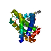 6u64C  6u65C  6u67C  2pqkS C: citing same article ( S: Starting model for refinement |
|---|---|
| Similar structure data |
-Links
 PDBj
PDBj

- Assembly
| Deposited unit | 
| ||||||||
|---|---|---|---|---|---|---|---|---|---|
| 1 | 
| ||||||||
| 2 | 
| ||||||||
| 3 | 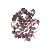
| ||||||||
| Unit cell |
|
-Components
| #1: Protein | Mass: 17838.229 Da / Num. of mol.: 3 Source method: isolated from a genetically manipulated source Source: (gene. exp.) Homo sapiens (human) / Gene: MCL1, BCL2L3 / Production host:  #2: Chemical | ChemComp-PZY /   Mass: 567.762 Da / Num. of mol.: 4 / Source method: obtained synthetically / Formula: C30H37N3O4S2 / Feature type: SUBJECT OF INVESTIGATION Mass: 567.762 Da / Num. of mol.: 4 / Source method: obtained synthetically / Formula: C30H37N3O4S2 / Feature type: SUBJECT OF INVESTIGATION#3: Water | ChemComp-HOH / |  Mass: 18.015 Da / Num. of mol.: 52 / Source method: isolated from a natural source / Formula: H2O Mass: 18.015 Da / Num. of mol.: 52 / Source method: isolated from a natural source / Formula: H2OHas ligand of interest | Y | |
|---|
-Experimental details
-Experiment
| Experiment | Method: X-RAY DIFFRACTION / Number of used crystals: 1 |
|---|
- Sample preparation
| Crystal | Density Matthews: 2.3 Å3/Da / Density % sol: 46.47 % |
|---|---|
| Crystal grow | Temperature: 293 K / Method: vapor diffusion, sitting drop Details: PEG3350 20-22.5%, 0.1 M Bis-Tris 6.5, 0.28 M Ammonium acetate |
-Data collection
| Diffraction | Mean temperature: 100 K / Serial crystal experiment: N | |||||||||||||||||||||||||||||||||||||||||||||||||||||||||||||||||||||||||||||||||||||||||||||||||||||||||||||||||||||||||||||||||||||||||||||||||||||||||||||||||||||||||||||||||||||||||||||
|---|---|---|---|---|---|---|---|---|---|---|---|---|---|---|---|---|---|---|---|---|---|---|---|---|---|---|---|---|---|---|---|---|---|---|---|---|---|---|---|---|---|---|---|---|---|---|---|---|---|---|---|---|---|---|---|---|---|---|---|---|---|---|---|---|---|---|---|---|---|---|---|---|---|---|---|---|---|---|---|---|---|---|---|---|---|---|---|---|---|---|---|---|---|---|---|---|---|---|---|---|---|---|---|---|---|---|---|---|---|---|---|---|---|---|---|---|---|---|---|---|---|---|---|---|---|---|---|---|---|---|---|---|---|---|---|---|---|---|---|---|---|---|---|---|---|---|---|---|---|---|---|---|---|---|---|---|---|---|---|---|---|---|---|---|---|---|---|---|---|---|---|---|---|---|---|---|---|---|---|---|---|---|---|---|---|---|---|---|---|---|
| Diffraction source | Source: SYNCHROTRON / Site: APS / Beamline: 21-ID-G / Wavelength: 0.97856 Å | |||||||||||||||||||||||||||||||||||||||||||||||||||||||||||||||||||||||||||||||||||||||||||||||||||||||||||||||||||||||||||||||||||||||||||||||||||||||||||||||||||||||||||||||||||||||||||||
| Detector | Type: MARRESEARCH / Detector: CCD / Date: Aug 15, 2019 | |||||||||||||||||||||||||||||||||||||||||||||||||||||||||||||||||||||||||||||||||||||||||||||||||||||||||||||||||||||||||||||||||||||||||||||||||||||||||||||||||||||||||||||||||||||||||||||
| Radiation | Protocol: SINGLE WAVELENGTH / Monochromatic (M) / Laue (L): M / Scattering type: x-ray | |||||||||||||||||||||||||||||||||||||||||||||||||||||||||||||||||||||||||||||||||||||||||||||||||||||||||||||||||||||||||||||||||||||||||||||||||||||||||||||||||||||||||||||||||||||||||||||
| Radiation wavelength | Wavelength: 0.97856 Å / Relative weight: 1 | |||||||||||||||||||||||||||||||||||||||||||||||||||||||||||||||||||||||||||||||||||||||||||||||||||||||||||||||||||||||||||||||||||||||||||||||||||||||||||||||||||||||||||||||||||||||||||||
| Reflection | Resolution: 2.9→200 Å / Num. obs: 12245 / % possible obs: 99.9 % / Redundancy: 18 % / Biso Wilson estimate: 71.33 Å2 / Rmerge(I) obs: 0.12 / Rpim(I) all: 0.028 / Rrim(I) all: 0.124 / Χ2: 1.095 / Net I/σ(I): 6.6 / Num. measured all: 220047 | |||||||||||||||||||||||||||||||||||||||||||||||||||||||||||||||||||||||||||||||||||||||||||||||||||||||||||||||||||||||||||||||||||||||||||||||||||||||||||||||||||||||||||||||||||||||||||||
| Reflection shell | Diffraction-ID: 1
|
- Processing
| Software |
| ||||||||||||||||||||||||||||||||||||||||||||||||||||||||||||||||||||||||||||||||||||||||||||||||||||||||||||
|---|---|---|---|---|---|---|---|---|---|---|---|---|---|---|---|---|---|---|---|---|---|---|---|---|---|---|---|---|---|---|---|---|---|---|---|---|---|---|---|---|---|---|---|---|---|---|---|---|---|---|---|---|---|---|---|---|---|---|---|---|---|---|---|---|---|---|---|---|---|---|---|---|---|---|---|---|---|---|---|---|---|---|---|---|---|---|---|---|---|---|---|---|---|---|---|---|---|---|---|---|---|---|---|---|---|---|---|---|---|
| Refinement | Method to determine structure: MOLECULAR REPLACEMENT Starting model: 2PQK Resolution: 2.9→36.07 Å / Cor.coef. Fo:Fc: 0.909 / Cor.coef. Fo:Fc free: 0.861 / Cross valid method: THROUGHOUT / σ(F): 0 / SU Rfree Blow DPI: 0.457
| ||||||||||||||||||||||||||||||||||||||||||||||||||||||||||||||||||||||||||||||||||||||||||||||||||||||||||||
| Displacement parameters | Biso max: 125.44 Å2 / Biso mean: 52.78 Å2 / Biso min: 24.15 Å2
| ||||||||||||||||||||||||||||||||||||||||||||||||||||||||||||||||||||||||||||||||||||||||||||||||||||||||||||
| Refine analyze | Luzzati coordinate error obs: 0.45 Å | ||||||||||||||||||||||||||||||||||||||||||||||||||||||||||||||||||||||||||||||||||||||||||||||||||||||||||||
| Refinement step | Cycle: final / Resolution: 2.9→36.07 Å
| ||||||||||||||||||||||||||||||||||||||||||||||||||||||||||||||||||||||||||||||||||||||||||||||||||||||||||||
| Refine LS restraints |
| ||||||||||||||||||||||||||||||||||||||||||||||||||||||||||||||||||||||||||||||||||||||||||||||||||||||||||||
| LS refinement shell | Resolution: 2.9→2.94 Å / Rfactor Rfree error: 0 / Total num. of bins used: 29
|
