[English] 日本語
Yorodumi
- PDB-4oq5: Crystal Structure of Human MCL-1 Bound to Inhibitor 4-(4-methylna... -

+
Open data


ID or keywords:

Loading...

-
Basic information

Entry
Database: PDB / ID: 4oq5
Title
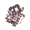
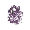
Crystal Structure of Human MCL-1 Bound to Inhibitor 4-(4-methylnaphthalen-1-yl)-2-{[(4-phenoxyphenyl)sulfonyl]amino}benzoic acid
ComponentsInduced myeloid leukemia cell differentiation protein Mcl-1
KeywordsAPOPTOSIS/INHIBITOR / APOPTOSIS-INHIBITOR complex
Function / homology
Function and homology information


positive regulation of oxidative stress-induced neuron intrinsic apoptotic signaling pathway / cellular homeostasis / cell fate determination / channel activity / mitochondrial fusion / Bcl-2 family protein complex / BH3 domain binding / negative regulation of anoikis / negative regulation of extrinsic apoptotic signaling pathway in absence of ligand / protein transmembrane transporter activity ...positive regulation of oxidative stress-induced neuron intrinsic apoptotic signaling pathway / cellular homeostasis / cell fate determination / channel activity / mitochondrial fusion / Bcl-2 family protein complex / BH3 domain binding / negative regulation of anoikis / negative regulation of extrinsic apoptotic signaling pathway in absence of ligand / protein transmembrane transporter activity / extrinsic apoptotic signaling pathway in absence of ligand / negative regulation of autophagy / release of cytochrome c from mitochondria / response to cytokine / intrinsic apoptotic signaling pathway in response to DNA damage / Signaling by ALK fusions and activated point mutants / regulation of apoptotic process / Interleukin-4 and Interleukin-13 signaling / mitochondrial outer membrane / positive regulation of apoptotic process / protein heterodimerization activity / DNA damage response / negative regulation of apoptotic process / protein homodimerization activity / mitochondrion / nucleoplasm / membrane / nucleus / cytoplasm / cytosol
Similarity search - Function
Apoptosis regulator, Mcl-1 / Blc2-like / Apoptosis Regulator Bcl-x / Apoptosis regulator, Bcl-2, BH3 motif, conserved site / Apoptosis regulator, Bcl-2 family BH3 motif signature. / Apoptosis regulator, Bcl-2, BH1 motif, conserved site / Apoptosis regulator, Bcl-2 family BH1 motif signature. / Apoptosis regulator, Bcl-2, BH2 motif, conserved site / Apoptosis regulator, Bcl-2 family BH2 motif signature. / BCL (B-Cell lymphoma); contains BH1, BH2 regions ...Apoptosis regulator, Mcl-1 / Blc2-like / Apoptosis Regulator Bcl-x / Apoptosis regulator, Bcl-2, BH3 motif, conserved site / Apoptosis regulator, Bcl-2 family BH3 motif signature. / Apoptosis regulator, Bcl-2, BH1 motif, conserved site / Apoptosis regulator, Bcl-2 family BH1 motif signature. / Apoptosis regulator, Bcl-2, BH2 motif, conserved site / Apoptosis regulator, Bcl-2 family BH2 motif signature. / BCL (B-Cell lymphoma); contains BH1, BH2 regions / Bcl-2 family / Bcl-2, Bcl-2 homology region 1-3 / Bcl2-like / Apoptosis regulator proteins, Bcl-2 family / BCL2-like apoptosis inhibitors family profile. / Bcl-2-like superfamily / Orthogonal Bundle / Mainly Alpha
Similarity search - Domain/homology
Chem-2UU / Induced myeloid leukemia cell differentiation protein Mcl-1
Similarity search - Component
Biological speciesHomo sapiens (human)
MethodX-RAY DIFFRACTION / SYNCHROTRON / MOLECULAR REPLACEMENT / Resolution: 2.86 Å
AuthorsPetros, A.M. / Swann, S.L. / Song, D. / Swinger, K. / Park, C. / Zhang, H. / Wendt, M.D. / Kunzer, A.R. / Souers, A.J. / Sun, C.
CitationJournal: Bioorg.Med.Chem.Lett. / Year: 2014
Title: Fragment-based discovery of potent inhibitors of the anti-apoptotic MCL-1 protein.
Authors: Petros, A.M. / Swann, S.L. / Song, D. / Swinger, K. / Park, C. / Zhang, H. / Wendt, M.D. / Kunzer, A.R. / Souers, A.J. / Sun, C.
History
DepositionFeb 7, 2014Deposition site: RCSB / Processing site: RCSB
Revision 1.0Mar 12, 2014Provider: repository / Type: Initial release
Revision 1.1Feb 28, 2024Group: Data collection / Database references / Derived calculations
Category: chem_comp_atom / chem_comp_bond ...chem_comp_atom / chem_comp_bond / database_2 / struct_ref_seq_dif / struct_site
Item: _database_2.pdbx_DOI / _database_2.pdbx_database_accession ..._database_2.pdbx_DOI / _database_2.pdbx_database_accession / _struct_ref_seq_dif.details / _struct_site.pdbx_auth_asym_id / _struct_site.pdbx_auth_comp_id / _struct_site.pdbx_auth_seq_id

-
Structure visualization

Structure viewerMolecule:
MolmilJmol/JSmol

Downloads & links

-
Assembly

Deposited unit
A: Induced myeloid leukemia cell differentiation protein Mcl-1
B: Induced myeloid leukemia cell differentiation protein Mcl-1
C: Induced myeloid leukemia cell differentiation protein Mcl-1
D: Induced myeloid leukemia cell differentiation protein Mcl-1
E: Induced myeloid leukemia cell differentiation protein Mcl-1
F: Induced myeloid leukemia cell differentiation protein Mcl-1
hetero molecules


Theoretical massNumber of molelcules
Total (without water)114,10012
Polymers111,0436
Non-polymers3,0576
Water2,774154
1
A: Induced myeloid leukemia cell differentiation protein Mcl-1
hetero molecules


Theoretical massNumber of molelcules
Total (without water)19,0172
Polymers18,5071
Non-polymers5101
Water181
TypeNameSymmetry operationNumber
identity operation1_555x,y,z1
2
B: Induced myeloid leukemia cell differentiation protein Mcl-1
hetero molecules


Theoretical massNumber of molelcules
Total (without water)19,0172
Polymers18,5071
Non-polymers5101
Water181
TypeNameSymmetry operationNumber
identity operation1_555x,y,z1
3
C: Induced myeloid leukemia cell differentiation protein Mcl-1
hetero molecules


Theoretical massNumber of molelcules
Total (without water)19,0172
Polymers18,5071
Non-polymers5101
Water181
TypeNameSymmetry operationNumber
identity operation1_555x,y,z1
4
D: Induced myeloid leukemia cell differentiation protein Mcl-1
hetero molecules


Theoretical massNumber of molelcules
Total (without water)19,0172
Polymers18,5071
Non-polymers5101
Water181
TypeNameSymmetry operationNumber
identity operation1_555x,y,z1
5
E: Induced myeloid leukemia cell differentiation protein Mcl-1
hetero molecules


Theoretical massNumber of molelcules
Total (without water)19,0172
Polymers18,5071
Non-polymers5101
Water181
TypeNameSymmetry operationNumber
identity operation1_555x,y,z1
6
F: Induced myeloid leukemia cell differentiation protein Mcl-1
hetero molecules


Theoretical massNumber of molelcules
Total (without water)19,0172
Polymers18,5071
Non-polymers5101
Water181
TypeNameSymmetry operationNumber
identity operation1_555x,y,z1
Unit cell
Length a, b, c (Å)71.112, 109.750, 76.544
Angle α, β, γ (deg.)90.00, 93.31, 90.00
Int Tables number4
Space group name H-MP1211

-
Components

#1: Protein
Induced myeloid leukemia cell differentiation protein Mcl-1 / Bcl-2-like protein 3 / Bcl2-L-3 / Bcl-2-related protein EAT/mcl1 / mcl1/EAT


Mass: 18507.146 Da / Num. of mol.: 6 / Fragment: UNP residues 174-326
Source method: isolated from a genetically manipulated source
Source: (gene. exp.) Homo sapiens (human) / Gene: MCL1, BCL2L3 / Production host: Escherichia coli (E. coli) / References: UniProt: Q07820
#2: Chemical
ChemComp-2UU / 4-(4-methylnaphthalen-1-yl)-2-{[(4-phenoxyphenyl)sulfonyl]amino}benzoic acid


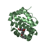
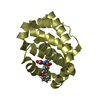
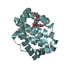
Mass: 509.572 Da / Num. of mol.: 6 / Source method: obtained synthetically / Formula: C30H23NO5S
#3: Water ChemComp-HOH / water


Mass: 18.015 Da / Num. of mol.: 154 / Source method: isolated from a natural source / Formula: H2O

-
Experimental details

-
Experiment

ExperimentMethod: X-RAY DIFFRACTION / Number of used crystals: 1

-
Sample preparation

CrystalDensity Matthews: 2.69 Å3/Da / Density % sol: 54.2 %
Crystal growTemperature: 277 K / Method: vapor diffusion, sitting drop / pH: 6.5
Details: 0.2 M ammonium sulfate, 0.1 M MES, 30% w/v PEG5000 MME, pH 6.5, VAPOR DIFFUSION, SITTING DROP, temperature 277K

-
Data collection

DiffractionMean temperature: 100 K
Diffraction sourceSource: SYNCHROTRON / Site: APS / Beamline: 17-ID / Wavelength: 1 Å
DetectorType: DECTRIS PILATUS 6M / Detector: PIXEL
RadiationProtocol: SINGLE WAVELENGTH / Monochromatic (M) / Laue (L): M / Scattering type: x-ray
Radiation wavelengthWavelength: 1 Å / Relative weight: 1
ReflectionResolution: 2.855→50 Å / Num. obs: 26737 / % possible obs: 99.9 % / Biso Wilson estimate: 73.89 Å2
Reflection shellHighest resolution: 2.855 Å

-
Processing

Software
NameVersionClassification
PHASERphasing
BUSTER2.11.4refinement
RefinementMethod to determine structure: MOLECULAR REPLACEMENT / Resolution: 2.86→48.15 Å / Cor.coef. Fo:Fc: 0.937 / Cor.coef. Fo:Fc free: 0.9125 / Cross valid method: THROUGHOUT / σ(F): 0 / SU Rfree Blow DPI: 0.354
RfactorNum. reflection% reflectionSelection details
Rfree0.2387 1340 5.01 %RANDOM
Rwork0.1975 ---
obs0.1996 26737 98.21 %-
Displacement parametersBiso mean: 60.49 Å2
Baniso -1Baniso -2Baniso -3
1--5.6398 Å20 Å21.3105 Å2
2--2.9896 Å20 Å2
3---2.6501 Å2
Refine analyzeLuzzati coordinate error obs: 0.348 Å
Refinement stepCycle: LAST / Resolution: 2.86→48.15 Å
ProteinNucleic acidLigandSolventTotal
Num. atoms7240 0 222 154 7616
Refine LS restraints
Refine-IDTypeDev idealNumberRestraint functionWeight
X-RAY DIFFRACTIONt_bond_d0.017728HARMONIC2
X-RAY DIFFRACTIONt_angle_deg1.1710480HARMONIC2
X-RAY DIFFRACTIONt_dihedral_angle_d2768SINUSOIDAL2
X-RAY DIFFRACTIONt_incorr_chiral_ct
X-RAY DIFFRACTIONt_pseud_angle
X-RAY DIFFRACTIONt_trig_c_planes180HARMONIC2
X-RAY DIFFRACTIONt_gen_planes1264HARMONIC5
X-RAY DIFFRACTIONt_it7728HARMONIC20
X-RAY DIFFRACTIONt_nbd
X-RAY DIFFRACTIONt_omega_torsion2.27
X-RAY DIFFRACTIONt_other_torsion21.12
X-RAY DIFFRACTIONt_improper_torsion
X-RAY DIFFRACTIONt_chiral_improper_torsion946SEMIHARMONIC5
X-RAY DIFFRACTIONt_sum_occupancies
X-RAY DIFFRACTIONt_utility_distance
X-RAY DIFFRACTIONt_utility_angle
X-RAY DIFFRACTIONt_utility_torsion
X-RAY DIFFRACTIONt_ideal_dist_contact9646SEMIHARMONIC4
LS refinement shellResolution: 2.86→2.98 Å / Total num. of bins used: 13
RfactorNum. reflection% reflection
Rfree0.2536 123 4.73 %
Rwork0.217 2478 -
all0.2187 2601 -
obs--98.21 %

+
About Yorodumi

-
News

-
Feb 9, 2022. New format data for meta-information of EMDB entries

New format data for meta-information of EMDB entries

  • Version 3 of the EMDB header file is now the official format.
  • The previous official version 1.9 will be removed from the archive.

Related info.:EMDB header

External links:wwPDB to switch to version 3 of the EMDB data model

-
Aug 12, 2020. Covid-19 info

Covid-19 info

URL: https://pdbjlvh1.pdbj.org/emnavi/covid19.php

New page: Covid-19 featured information page in EM Navigator.

Related info.:Covid-19 info / Mar 5, 2020. Novel coronavirus structure data

+
Mar 5, 2020. Novel coronavirus structure data

Novel coronavirus structure data

Related info.:Yorodumi Speices / Aug 12, 2020. Covid-19 info

External links:COVID-19 featured content - PDBj / Molecule of the Month (242):Coronavirus Proteases

+
Jan 31, 2019. EMDB accession codes are about to change! (news from PDBe EMDB page)

EMDB accession codes are about to change! (news from PDBe EMDB page)

  • The allocation of 4 digits for EMDB accession codes will soon come to an end. Whilst these codes will remain in use, new EMDB accession codes will include an additional digit and will expand incrementally as the available range of codes is exhausted. The current 4-digit format prefixed with “EMD-” (i.e. EMD-XXXX) will advance to a 5-digit format (i.e. EMD-XXXXX), and so on. It is currently estimated that the 4-digit codes will be depleted around Spring 2019, at which point the 5-digit format will come into force.
  • The EM Navigator/Yorodumi systems omit the EMD- prefix.

Related info.:Q: What is EMD? / ID/Accession-code notation in Yorodumi/EM Navigator

External links:EMDB Accession Codes are Changing Soon! / Contact to PDBj

+
Jul 12, 2017. Major update of PDB

Major update of PDB

  • wwPDB released updated PDB data conforming to the new PDBx/mmCIF dictionary.
  • This is a major update changing the version number from 4 to 5, and with Remediation, in which all the entries are updated.
  • In this update, many items about electron microscopy experimental information are reorganized (e.g. em_software).
  • Now, EM Navigator and Yorodumi are based on the updated data.

External links:wwPDB Remediation / Enriched Model Files Conforming to OneDep Data Standards Now Available in the PDB FTP Archive

-
Yorodumi

Thousand views of thousand structures

  • Yorodumi is a browser for structure data from EMDB, PDB, SASBDB, etc.
  • This page is also the successor to EM Navigator detail page, and also detail information page/front-end page for Omokage search.
  • The word "yorodu" (or yorozu) is an old Japanese word meaning "ten thousand". "mi" (miru) is to see.

Related info.:EMDB / PDB / SASBDB / Comparison of 3 databanks / Yorodumi Search / Aug 31, 2016. New EM Navigator & Yorodumi / Yorodumi Papers / Jmol/JSmol / Function and homology information / Changes in new EM Navigator and Yorodumi

Read more