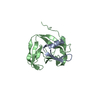
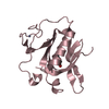
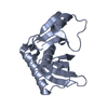
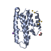
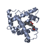
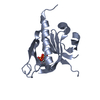
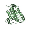
- PDB-2uzp: Crystal structure of the C2 domain of human protein kinase C gamma. -
+
Open data
ID or keywords:
Loading...
-
Basic information
Entry
Database: PDB / ID: 2uzp
Title
Crystal structure of the C2 domain of human protein kinase C gamma.
Components
PROTEIN KINASE C GAMMA TYPE
Keywords
TRANSFERASE / METAL-BINDING / KINASE / ZINC-FINGER / ATP-BINDING / SPINOCEREBELLAR ATAXIA / CALCIUM-BINDING PROTEIN / SERINE/THREONINE-PROTEIN KINASE / PHOSPHOLIPID BINDING PROTEIN / NUCLEOTIDE-BINDING / PHORBOL- ESTER BINDING / PHOSPHORYLATION / PROTEIN KINASE C / NEURODEGENERATION
Function / homology
Function and homology information
regulation of response to food / Disinhibition of SNARE formation / Response to elevated platelet cytosolic Ca2+ / calcium,diacylglycerol-dependent serine/threonine kinase activity / positive regulation of mismatch repair / Trafficking of GluR2-containing AMPA receptors / WNT5A-dependent internalization of FZD4 / protein kinase C / negative regulation of proteasomal protein catabolic process / diacylglycerol-dependent serine/threonine kinase activity ...regulation of response to food / Disinhibition of SNARE formation / Response to elevated platelet cytosolic Ca2+ / calcium,diacylglycerol-dependent serine/threonine kinase activity / positive regulation of mismatch repair / Trafficking of GluR2-containing AMPA receptors / WNT5A-dependent internalization of FZD4 / protein kinase C / negative regulation of proteasomal protein catabolic process / diacylglycerol-dependent serine/threonine kinase activity / response to angiotensin / innervation / Calmodulin induced events / response to pain / response to psychosocial stress / response to morphine / regulation of synaptic vesicle exocytosis / postsynaptic cytosol / presynaptic cytosol / protein serine/threonine/tyrosine kinase activity / negative regulation of protein ubiquitination / calyx of Held / synaptic membrane / negative regulation of protein catabolic process / regulation of circadian rhythm / response to toxic substance / cell-cell junction / long-term synaptic potentiation / rhythmic process / G alpha (z) signalling events / phospholipase C-activating G protein-coupled receptor signaling pathway / chemical synaptic transmission / negative regulation of neuron apoptotic process / learning or memory / protein kinase activity / postsynaptic density / intracellular signal transduction / protein serine kinase activity / protein serine/threonine kinase activity / dendrite / perinuclear region of cytoplasm / zinc ion binding / ATP binding / nucleus / plasma membrane / cytosol Similarity search - Function
Protein kinase C, alpha/beta/gamma types / Protein kinase, C-terminal / Protein kinase C terminal domain / Diacylglycerol/phorbol-ester binding / Phorbol esters/diacylglycerol binding domain (C1 domain) / C2 domain / Zinc finger phorbol-ester/DAG-type signature. / Protein kinase C conserved region 2 (CalB) / C2 domain / Zinc finger phorbol-ester/DAG-type profile. ...Protein kinase C, alpha/beta/gamma types / Protein kinase, C-terminal / Protein kinase C terminal domain / Diacylglycerol/phorbol-ester binding / Phorbol esters/diacylglycerol binding domain (C1 domain) / C2 domain / Zinc finger phorbol-ester/DAG-type signature. / Protein kinase C conserved region 2 (CalB) / C2 domain / Zinc finger phorbol-ester/DAG-type profile. / Protein kinase C conserved region 1 (C1) domains (Cysteine-rich domains) / Protein kinase C-like, phorbol ester/diacylglycerol-binding domain / C2 domain / C2 domain profile. / C1-like domain superfamily / Extension to Ser/Thr-type protein kinases / AGC-kinase, C-terminal / AGC-kinase C-terminal domain profile. / C2 domain superfamily / Serine/threonine-protein kinase, active site / Serine/Threonine protein kinases active-site signature. / Protein kinase domain / Serine/Threonine protein kinases, catalytic domain / Protein kinase, ATP binding site / Protein kinases ATP-binding region signature. / Protein kinase domain profile. / Protein kinase domain / Protein kinase-like domain superfamily / Immunoglobulin-like / Sandwich / Mainly Beta Similarity search - Domain/homology
PROTEINKINASECGAMMATYPE / PROTEIN KINASE C GAMMA / PKC-GAMMA
Mass: 16533.654 Da / Num. of mol.: 3 / Fragment: C2 DOMAIN, RESIDUES 154-295 Source method: isolated from a genetically manipulated source Details: SCHIFFS BASE LINK BETWEEN SER -1 AND PYRIDOXAL PHOSPHATE PLP300 Source: (gene. exp.) HOMO SAPIENS (human) / Plasmid: PNIC28-BSA4 / Production host: ESCHERICHIA COLI (E. coli) / Strain (production host): BL21(DE3) / References: UniProt: P05129, protein kinase C
Resolution: 2→50 Å / Cor.coef. Fo:Fc: 0.961 / Cor.coef. Fo:Fc free: 0.947 / SU B: 5.117 / SU ML: 0.088 / TLS residual ADP flag: LIKELY RESIDUAL / Cross valid method: THROUGHOUT / ESU R: 0.135 / ESU R Free: 0.128 / Stereochemistry target values: MAXIMUM LIKELIHOOD Details: HYDROGENS HAVE BEEN ADDED IN THE RIDING POSITIONS. AMINO TERMINUS IS MODIFIED BY PYRIXODAL PHOSPHATE DURING CRYSTALLIZATION AND TIGHTLY COORDINATES A COBALT ION
Rfactor
Num. reflection
% reflection
Selection details
Rfree
0.205
2295
5 %
RANDOM
Rwork
0.171
-
-
-
obs
0.172
43180
100 %
-
Solvent computation
Ion probe radii: 0.8 Å / Shrinkage radii: 0.8 Å / VDW probe radii: 1.4 Å / Solvent model: MASK
 Movie
Movie Controller

 Yorodumi
 Open data
 Basic information
 Components
 Keywords
 Function and homology information
 HOMO SAPIENS (human)
 X-RAY DIFFRACTION /
  Authors
 Citation
 Structure visualization
 Downloads & links
 Other downloads
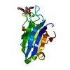


 PDBj
PDBj


 Assembly
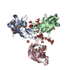

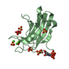
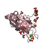


 Mass: 247.142 Da / Num. of mol.: 3 / Source method: obtained synthetically / Formula: C8H10NO6P
 Mass: 58.933 Da / Num. of mol.: 3 / Source method: obtained synthetically / Formula: Co
 Mass: 40.078 Da / Num. of mol.: 9 / Source method: obtained synthetically / Formula: Ca
 Mass: 96.063 Da / Num. of mol.: 20 / Source method: obtained synthetically / Formula: SO4
 Mass: 62.068 Da / Num. of mol.: 8 / Source method: obtained synthetically / Formula: C2H6O2
 Sample preparation
 / Beamline: X10SA / Wavelength: 0.9999
  Processing