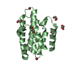
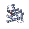
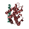
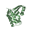
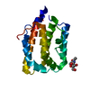
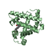
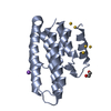
- PDB-4acl: 3D Structure of DotU from Francisella novicida -
+
Open data
ID or keywords:
Loading...
-
Basic information
Entry
Database: PDB / ID: 4acl
Title
3D Structure of DotU from Francisella novicida
Components
TSSL
Keywords
PROTEIN TRANSPORT / T6SS / MICROBIAL SURFACE STRUCTURES / MEMBRANE PROTEIN / VIRULENCE FACTOR
Function / homology
Type IV / VI secretion system, DotU / Type IV / VI secretion system, DotU / Type IV / VI secretion system, DotU superfamily / Type VI secretion system protein DotU / Serine Threonine Protein Phosphatase 5, Tetratricopeptide repeat / Alpha Horseshoe / Mainly Alpha / : / Type IV / VI secretion system DotU domain-containing protein
 Movie
Movie Controller


 Open data
 Basic information
 Components
 Keywords
 Function and homology information
 FRANCISELLA NOVICIDA (bacteria)
 X-RAY DIFFRACTION /
  Authors
 Citation
 Structure visualization
 Downloads & links
 Other downloads


 PDBj
PDBj

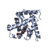
 Assembly
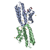

 Mass: 22.990 Da / Num. of mol.: 1 / Source method: obtained synthetically / Formula: Na
 Mass: 62.068 Da / Num. of mol.: 2 / Source method: obtained synthetically / Formula: C2H6O2
 Mass: 196.967 Da / Num. of mol.: 12 / Source method: obtained synthetically / Formula: Au
 Mass: 18.015 Da / Num. of mol.: 40 / Source method: isolated from a natural source / Formula: H2O
 Sample preparation
 Processing