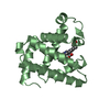
 Movie
Movie Controller


+ Open data

- Basic information
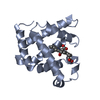
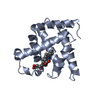
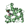
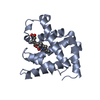
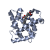
| Entry | Database: PDB / ID: 1mwd | ||||||
|---|---|---|---|---|---|---|---|
| Title | WILD TYPE DEOXY MYOGLOBIN | ||||||
 Components Components | PROTEIN (MYOGLOBIN) | ||||||
 Keywords Keywords | OXYGEN STORAGE/TRANSPORT / OXYGEN STORAGE / DEOXY MYOGLOBIN / OXYGEN STORAGE-TRANSPORT COMPLEX | ||||||
| Function / homology |  Function and homology information Function and homology informationIntracellular oxygen transport / Oxidoreductases; Acting on other nitrogenous compounds as donors / nitrite reductase activity / oxygen transport / sarcoplasm / Oxidoreductases; Acting on a peroxide as acceptor; Peroxidases / skeletal muscle contraction / removal of superoxide radicals / oxygen carrier activity / peroxidase activity ...Intracellular oxygen transport / Oxidoreductases; Acting on other nitrogenous compounds as donors / nitrite reductase activity / oxygen transport / sarcoplasm / Oxidoreductases; Acting on a peroxide as acceptor; Peroxidases / skeletal muscle contraction / removal of superoxide radicals / oxygen carrier activity / peroxidase activity / oxygen binding / heme binding / metal ion binding Similarity search - Function | ||||||
| Biological species |  | ||||||
| Method |  X-RAY DIFFRACTION / OTHER / Resolution: 1.8 Å X-RAY DIFFRACTION / OTHER / Resolution: 1.8 Å | ||||||
 Authors Authors | Murshudov, G.N. / Krzywda, S. / Brzozowski, A.M. / Jaskolski, M. / Scott, E.E. / Klizas, A.A. / Gibson, Q.H. / Olson, J.S. / Wilkinson, A.J. | ||||||
 Citation Citation | Journal: Biochemistry / Year: 1998 Title: Stabilizing bound O2 in myoglobin by valine68 (E11) to asparagine substitution. Authors: Krzywda, S. / Murshudov, G.N. / Brzozowski, A.M. / Jaskolski, M. / Scott, E.E. / Klizas, S.A. / Gibson, Q.H. / Olson, J.S. / Wilkinson, A.J. | ||||||
| History |
|
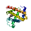
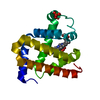
- Structure visualization
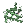
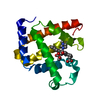
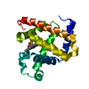
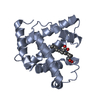
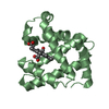
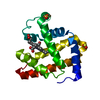
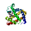
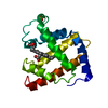
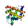
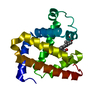
| Structure viewer | Molecule: MolmilJmol/JSmol |
|---|
- Downloads & links
-Download
| PDBx/mmCIF format | 1mwd.cif.gz | 85.2 KB | Display | PDBx/mmCIF format |
|---|---|---|---|---|
| PDB format | pdb1mwd.ent.gz | 65.6 KB | Display | PDB format |
| PDBx/mmJSON format | 1mwd.json.gz | Tree view | PDBx/mmJSON format | |
| Others |  Other downloads Other downloads |
-Validation report
| Arichive directory | https://data.pdbj.org/pub/pdb/validation_reports/mw/1mwdftp://data.pdbj.org/pub/pdb/validation_reports/mw/1mwd | HTTPS FTP |
|---|
-Related structure data


-Links
 PDBj
PDBj


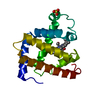
- Assembly
| Deposited unit | 
| ||||||||
|---|---|---|---|---|---|---|---|---|---|
| 1 | 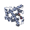
| ||||||||
| 2 | 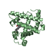
| ||||||||
| Unit cell |
|
-Components
| #1: Protein | Mass: 16983.445 Da / Num. of mol.: 2 Source method: isolated from a genetically manipulated source Details: MYOGLOBIN / Source: (gene. exp.)  #2: Chemical |   Mass: 616.487 Da / Num. of mol.: 2 / Source method: obtained synthetically / Formula: C34H32FeN4O4 Mass: 616.487 Da / Num. of mol.: 2 / Source method: obtained synthetically / Formula: C34H32FeN4O4#3: Water | ChemComp-HOH / |  Mass: 18.015 Da / Num. of mol.: 456 / Source method: isolated from a natural source / Formula: H2O Mass: 18.015 Da / Num. of mol.: 456 / Source method: isolated from a natural source / Formula: H2O |
|---|
-Experimental details
-Experiment
| Experiment | Method: X-RAY DIFFRACTION / Number of used crystals: 1 |
|---|
- Sample preparation
| Crystal | Density Matthews: 3.46 Å3/Da / Density % sol: 50 % | ||||||||||||||||||||
|---|---|---|---|---|---|---|---|---|---|---|---|---|---|---|---|---|---|---|---|---|---|
| Crystal grow | pH: 7.1 Details: 0.1 PHOSPHATE BUFFER 72-80% SATURATED AMMONIUM SULPHATE, pH 7.1 | ||||||||||||||||||||
| Crystal | *PLUS | ||||||||||||||||||||
| Crystal grow | *PLUS Method: vapor diffusion, hanging drop | ||||||||||||||||||||
| Components of the solutions | *PLUS
|
-Data collection
| Diffraction | Mean temperature: 150 K |
|---|---|
| Diffraction source | Source: ROTATING ANODE / Type: RIGAKU RU200 / Wavelength: 1.5418 |
| Detector | Type: RIGAKU RAXIS / Detector: IMAGE PLATE |
| Radiation | Protocol: SINGLE WAVELENGTH / Monochromatic (M) / Laue (L): M / Scattering type: x-ray |
| Radiation wavelength | Wavelength: 1.5418 Å / Relative weight: 1 |
| Reflection | Resolution: 1.8→24 Å / Num. obs: 42068 / % possible obs: 96.5 % / Redundancy: 7.9 % / Biso Wilson estimate: 26.5 Å2 / Rsym value: 0.04 / Net I/σ(I): 21.4 |
| Reflection | *PLUS Highest resolution: 1.8 Å / Rmerge(I) obs: 0.04 |
| Reflection shell | *PLUS Rmerge(I) obs: 0.291 |
- Processing
| Software |
| ||||||||||||||||||||||||||||||||||||||||||||||||||||||||||||||||||||||||||||||||||||
|---|---|---|---|---|---|---|---|---|---|---|---|---|---|---|---|---|---|---|---|---|---|---|---|---|---|---|---|---|---|---|---|---|---|---|---|---|---|---|---|---|---|---|---|---|---|---|---|---|---|---|---|---|---|---|---|---|---|---|---|---|---|---|---|---|---|---|---|---|---|---|---|---|---|---|---|---|---|---|---|---|---|---|---|---|---|
| Refinement | Method to determine structure: OTHER / Resolution: 1.8→24 Å / SU B: 2.58 / Cross valid method: THROUGHOUT / σ(F): 0
| ||||||||||||||||||||||||||||||||||||||||||||||||||||||||||||||||||||||||||||||||||||
| Displacement parameters | Biso mean: 31 Å2
| ||||||||||||||||||||||||||||||||||||||||||||||||||||||||||||||||||||||||||||||||||||
| Refinement step | Cycle: LAST / Resolution: 1.8→24 Å
| ||||||||||||||||||||||||||||||||||||||||||||||||||||||||||||||||||||||||||||||||||||
| Refine LS restraints |
| ||||||||||||||||||||||||||||||||||||||||||||||||||||||||||||||||||||||||||||||||||||
| Software | *PLUS Name: REFMAC / Classification: refinement | ||||||||||||||||||||||||||||||||||||||||||||||||||||||||||||||||||||||||||||||||||||
| Refinement | *PLUS Highest resolution: 1.8 Å / σ(F): 0 / % reflection Rfree: 5 % / Rfactor obs: 0.196 | ||||||||||||||||||||||||||||||||||||||||||||||||||||||||||||||||||||||||||||||||||||
| Solvent computation | *PLUS | ||||||||||||||||||||||||||||||||||||||||||||||||||||||||||||||||||||||||||||||||||||
| Displacement parameters | *PLUS Biso mean: 31 Å2 | ||||||||||||||||||||||||||||||||||||||||||||||||||||||||||||||||||||||||||||||||||||
| Refine LS restraints | *PLUS
|
