 Movie
Movie Controller

[English] 日本語
 Yorodumi
- PDB-1ycb: DISTAL POCKET POLARITY IN LIGAND BINDING TO MYOGLOBIN: DEOXY AND ... -
+ Open data

- Basic information
| Entry | Database: PDB / ID: 1ycb | ||||||
|---|---|---|---|---|---|---|---|
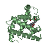
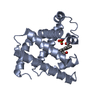
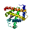
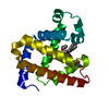
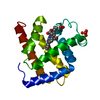
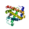
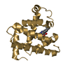
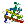
| Title | DISTAL POCKET POLARITY IN LIGAND BINDING TO MYOGLOBIN: DEOXY AND CARBONMONOXY FORMS OF A THREONINE68 (E11) MUTANT INVESTIGATED BY X-RAY CRYSTALLOGRAPHY AND INFRARED SPECTROSCOPY | ||||||
 Components Components | MYOGLOBIN | ||||||
 Keywords Keywords | OXYGEN STORAGE | ||||||
| Function / homology |  Function and homology information Function and homology informationIntracellular oxygen transport / Oxidoreductases; Acting on other nitrogenous compounds as donors / nitrite reductase activity / oxygen transport / sarcoplasm / Oxidoreductases; Acting on a peroxide as acceptor; Peroxidases / skeletal muscle contraction / removal of superoxide radicals / oxygen carrier activity / peroxidase activity ...Intracellular oxygen transport / Oxidoreductases; Acting on other nitrogenous compounds as donors / nitrite reductase activity / oxygen transport / sarcoplasm / Oxidoreductases; Acting on a peroxide as acceptor; Peroxidases / skeletal muscle contraction / removal of superoxide radicals / oxygen carrier activity / peroxidase activity / oxygen binding / heme binding / metal ion binding Similarity search - Function | ||||||
| Biological species |  | ||||||
| Method |  X-RAY DIFFRACTION / Resolution: 2.1 Å X-RAY DIFFRACTION / Resolution: 2.1 Å | ||||||
 Authors Authors | Cameron, A.D. / Smerdon, S.J. / Wilkinson, A.J. / Habash, J. / Helliwell, J.R. | ||||||
 Citation Citation | Journal: Biochemistry / Year: 1993 Title: Distal pocket polarity in ligand binding to myoglobin: deoxy and carbonmonoxy forms of a threonine68(E11) mutant investigated by X-ray crystallography and infrared spectroscopy. Authors: Cameron, A.D. / Smerdon, S.J. / Wilkinson, A.J. / Habash, J. / Helliwell, J.R. / Li, T. / Olson, J.S. #1: Journal: Biochemistry / Year: 1992Title: High Resolution X-Ray Structures of Pig Metmyoglobin and Two Cd3 Mutants Mb(Lys45-> Arg) and Mb(Lys45-> Ser) Authors: Oldfield, T.J. / Smerdon, S.J. / Dauter, Z. / Petratos, K. / Wilson, K.S. / Wilkinson, A.J. #2: Journal: Biochemistry / Year: 1991Title: Distal Polarity in Ligand Binding to Myoglobin: Structural and Functional Characterization of a Threonine68(E11) Mutant Authors: Smerdon, S.J. / Dodson, G.G. / Wilkinson, A.J. / Gibson, Q.H. / Blackmore, R.S. / Carver, T.E. / Olson, J.S. #3: Journal: Acta Crystallogr.,Sect.B / Year: 1990Title: Determination of the Crystal Structure of Recombinant Pig Myoglobin by Molecular Replacement and its Refinement Authors: Smerdon, S.J. / Oldfield, T.J. / Dodson, E.J. / Dodson, G.G. / Hubbard, R.E. / Wilkinson, A.J. #4: Journal: Protein Eng. / Year: 1988Title: Apomyoglobin as a Molecular Recognition Surface: Expression, Reconstitution and Crystallisation of Recombinant Porcine Myoglobin in Escherichia Coli Authors: Dodson, G.G. / Hubbard, R.E. / Oldfield, T.J. / Smerdon, S.J. / Wilkinson, A.J. | ||||||
| History |
|
- Structure visualization
| Structure viewer | Molecule: MolmilJmol/JSmol |
|---|
- Downloads & links
-Download
| PDBx/mmCIF format | 1ycb.cif.gz | 77.5 KB | Display | PDBx/mmCIF format |
|---|---|---|---|---|
| PDB format | pdb1ycb.ent.gz | 58.6 KB | Display | PDB format |
| PDBx/mmJSON format | 1ycb.json.gz | Tree view | PDBx/mmJSON format | |
| Others |  Other downloads Other downloads |
-Validation report
| Arichive directory | https://data.pdbj.org/pub/pdb/validation_reports/yc/1ycbftp://data.pdbj.org/pub/pdb/validation_reports/yc/1ycb | HTTPS FTP |
|---|
-Related structure data


-Links
 PDBj
PDBj


- Assembly
| Deposited unit | 
| ||||||||
|---|---|---|---|---|---|---|---|---|---|
| 1 | 
| ||||||||
| 2 | 
| ||||||||
| Unit cell |
| ||||||||
| Atom site foot note | 1: PHE A 151 - GLN A 152 OMEGA ANGLE = 146.397 PEPTIDE BOND DEVIATES SIGNIFICANTLY FROM TRANS CONFORMATION |
-Components
| #1: Protein | Mass: 16985.418 Da / Num. of mol.: 2 Source method: isolated from a genetically manipulated source Source: (gene. exp.) #2: Chemical |   Mass: 616.487 Da / Num. of mol.: 2 / Source method: obtained synthetically / Formula: C34H32FeN4O4 Mass: 616.487 Da / Num. of mol.: 2 / Source method: obtained synthetically / Formula: C34H32FeN4O4#3: Water | ChemComp-HOH / |  Mass: 18.015 Da / Num. of mol.: 109 / Source method: isolated from a natural source / Formula: H2O Mass: 18.015 Da / Num. of mol.: 109 / Source method: isolated from a natural source / Formula: H2O |
|---|
-Experimental details
-Experiment
| Experiment | Method: X-RAY DIFFRACTION |
|---|
- Sample preparation
| Crystal grow | *PLUS pH: 7.1 / Method: vapor diffusion, hanging drop / Details: under 1 atm of argon | ||||||||||||||||||||||||||||||
|---|---|---|---|---|---|---|---|---|---|---|---|---|---|---|---|---|---|---|---|---|---|---|---|---|---|---|---|---|---|---|---|
| Components of the solutions | *PLUS
|
-Data collection
| Reflection | *PLUS Highest resolution: 2.1 Å / Num. obs: 18251 / Rmerge(I) obs: 0.111 |
|---|
- Processing
| Software | Name: PROLSQ / Classification: refinement | ||||||||||||||||||||||||||||||||||||||||||||||||||||||||||||||||||||||||||||||||||||
|---|---|---|---|---|---|---|---|---|---|---|---|---|---|---|---|---|---|---|---|---|---|---|---|---|---|---|---|---|---|---|---|---|---|---|---|---|---|---|---|---|---|---|---|---|---|---|---|---|---|---|---|---|---|---|---|---|---|---|---|---|---|---|---|---|---|---|---|---|---|---|---|---|---|---|---|---|---|---|---|---|---|---|---|---|---|
| Refinement | Resolution: 2.1→8 Å / σ(F): 0 Details: THIS STRUCTURE WAS REFINED AGAINST LAUE DATA TO 2.1 ANGSTROMS RESOLUTION. THE DATA SET IS 63% COMPLETE OVERALL BUT ONLY 38% OF REFLECTIONS ARE PRESENT BELOW 2DMIN. AS A RESULT THE ELECTRON ...Details: THIS STRUCTURE WAS REFINED AGAINST LAUE DATA TO 2.1 ANGSTROMS RESOLUTION. THE DATA SET IS 63% COMPLETE OVERALL BUT ONLY 38% OF REFLECTIONS ARE PRESENT BELOW 2DMIN. AS A RESULT THE ELECTRON DENSITY MAPS HAVE BREAKS PERIODICALLY ALONG THE MAIN CHAIN. THOSE ATOMS WHICH COULD NOT BE OBSERVED IN ANY MAP HAVE BEEN ASSIGNED AN OCCUPANCY OF 0.0. DURING THE COURSE OF REFINEMENT REASONABLY TIGHT RESTRAINTS WERE APPLIED TO THE GEOMETRY WHILE MOST ATTENTION WAS PAID TO THE HAEM GROUP AND ITS ENVIRONMENT. THIS STRATEGY WAS ADOPTED AS THE PURPOSE OF THE STUDY WAS TO DEFINE THE LOCATION OF WATER MOLECULES IN THE DISTAL POCKET.
| ||||||||||||||||||||||||||||||||||||||||||||||||||||||||||||||||||||||||||||||||||||
| Refinement step | Cycle: LAST / Resolution: 2.1→8 Å
| ||||||||||||||||||||||||||||||||||||||||||||||||||||||||||||||||||||||||||||||||||||
| Refine LS restraints |
| ||||||||||||||||||||||||||||||||||||||||||||||||||||||||||||||||||||||||||||||||||||
| Software | *PLUS Name: PROLSQ / Classification: refinement | ||||||||||||||||||||||||||||||||||||||||||||||||||||||||||||||||||||||||||||||||||||
| Refinement | *PLUS Highest resolution: 2.1 Å / Lowest resolution: 8 Å / Num. reflection all: 18200 / σ(F): 0 / Rfactor all: 0.198 | ||||||||||||||||||||||||||||||||||||||||||||||||||||||||||||||||||||||||||||||||||||
| Solvent computation | *PLUS | ||||||||||||||||||||||||||||||||||||||||||||||||||||||||||||||||||||||||||||||||||||
| Displacement parameters | *PLUS |
