 Movie
Movie Controller

[English] 日本語
 Yorodumi
- PDB-1dsy: C2 DOMAIN FROM PROTEIN KINASE C (ALPHA) COMPLEXED WITH CA2+ AND P... -
+ Open data

- Basic information
| Entry | Database: PDB / ID: 1dsy | ||||||
|---|---|---|---|---|---|---|---|
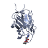
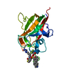
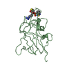
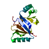
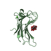
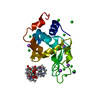
| Title | C2 DOMAIN FROM PROTEIN KINASE C (ALPHA) COMPLEXED WITH CA2+ AND PHOSPHATIDYLSERINE | ||||||
 Components Components | PROTEIN KINASE C, ALPHA TYPE | ||||||
 Keywords Keywords | TRANSFERASE / CALCIUM++ / PHOSPHOLIPID BINDING PROTEIN / CALCIUM-BINDING PROTEIN / PHOSPHATIDYLSERINE / PROTEIN KINASE C | ||||||
| Function / homology |  Function and homology information Function and homology informationEGFR Transactivation by Gastrin / HuR (ELAVL1) binds and stabilizes mRNA / Acetylcholine regulates insulin secretion / Depolymerization of the Nuclear Lamina / Signaling by SCF-KIT / Regulation of KIT signaling / Calmodulin induced events / Disinhibition of SNARE formation / SHC1 events in ERBB2 signaling / Response to elevated platelet cytosolic Ca2+ ...EGFR Transactivation by Gastrin / HuR (ELAVL1) binds and stabilizes mRNA / Acetylcholine regulates insulin secretion / Depolymerization of the Nuclear Lamina / Signaling by SCF-KIT / Regulation of KIT signaling / Calmodulin induced events / Disinhibition of SNARE formation / SHC1 events in ERBB2 signaling / Response to elevated platelet cytosolic Ca2+ / Syndecan interactions / response to phorbol 13-acetate 12-myristate / positive regulation of dense core granule biogenesis / cone photoreceptor outer segment / VEGFR2 mediated cell proliferation / positive regulation of angiotensin-activated signaling pathway / calcium,diacylglycerol-dependent serine/threonine kinase activity / RET signaling / histone H3T6 kinase activity / protein kinase C signaling / ooplasm / central nervous system neuron axonogenesis / desmosome assembly / WNT5A-dependent internalization of FZD4 / RHO GTPases Activate NADPH Oxidases / protein kinase C / cellular response to carbohydrate stimulus / diacylglycerol-dependent serine/threonine kinase activity / regulation of platelet aggregation / negative regulation of glial cell apoptotic process / renal sodium ion absorption / negative regulation of D-glucose import across plasma membrane / regulation of muscle contraction / positive regulation of macrophage differentiation / induction of positive chemotaxis / alphav-beta3 integrin-PKCalpha complex / regulation of the force of heart contraction / signal transduction involved in regulation of gene expression / positive regulation of lipopolysaccharide-mediated signaling pathway / positive regulation of bone resorption / positive regulation of cytokine production involved in inflammatory response / positive regulation of synapse assembly / muscle cell cellular homeostasis / response to corticosterone / positive regulation of cardiac muscle hypertrophy / regulation of synaptic vesicle exocytosis / Trafficking of GluR2-containing AMPA receptors / positive regulation of exocytosis / intercalated disc / positive regulation of blood vessel endothelial cell migration / chondrocyte differentiation / response to mechanical stimulus / presynaptic cytosol / negative regulation of MAPK cascade / neutrophil chemotaxis / positive regulation of endothelial cell proliferation / negative regulation of insulin receptor signaling pathway / negative regulation of cytokine production involved in inflammatory response / positive regulation of mitotic cell cycle / positive regulation of endothelial cell migration / intrinsic apoptotic signaling pathway / positive regulation of cell adhesion / positive regulation of smooth muscle cell proliferation / response to interleukin-1 / calyx of Held / presynaptic modulation of chemical synaptic transmission / stem cell differentiation / response to reactive oxygen species / establishment of protein localization / phosphatidylinositol 3-kinase/protein kinase B signal transduction / integrin binding / response to peptide hormone / mitochondrial membrane / response to toxic substance / intracellular calcium ion homeostasis / apical part of cell / positive regulation of angiogenesis / positive regulation of inflammatory response / response to estradiol / angiogenesis / response to ethanol / proteasome-mediated ubiquitin-dependent protein catabolic process / learning or memory / protein kinase activity / positive regulation of ERK1 and ERK2 cascade / positive regulation of phosphatidylinositol 3-kinase/protein kinase B signal transduction / cell adhesion / negative regulation of translation / intracellular signal transduction / positive regulation of cell migration / ciliary basal body / signaling receptor binding / negative regulation of cell population proliferation / protein serine kinase activity / axon / protein serine/threonine kinase activity / neuronal cell body / lipid binding / dendrite / perinuclear region of cytoplasm Similarity search - Function | ||||||
| Biological species |  | ||||||
| Method |  X-RAY DIFFRACTION / Resolution: 2.6 Å X-RAY DIFFRACTION / Resolution: 2.6 Å | ||||||
 Authors Authors | Verdaguer, N. / Corbalan-Garcia, S. / Ochoa, W.F. / Fita, I. / Gomez-Fernandez, J.C. | ||||||
 Citation Citation | Journal: EMBO J. / Year: 1999 Title: Ca(2+) bridges the C2 membrane-binding domain of protein kinase Calpha directly to phosphatidylserine. Authors: Verdaguer, N. / Corbalan-Garcia, S. / Ochoa, W.F. / Fita, I. / Gomez-Fernandez, J.C. | ||||||
| History |
|
- Structure visualization
| Structure viewer | Molecule: MolmilJmol/JSmol |
|---|
- Downloads & links
-Download
| PDBx/mmCIF format | 1dsy.cif.gz | 43.9 KB | Display | PDBx/mmCIF format |
|---|---|---|---|---|
| PDB format | pdb1dsy.ent.gz | 30.1 KB | Display | PDB format |
| PDBx/mmJSON format | 1dsy.json.gz | Tree view | PDBx/mmJSON format | |
| Others |  Other downloads Other downloads |
-Validation report
| Arichive directory | https://data.pdbj.org/pub/pdb/validation_reports/ds/1dsyftp://data.pdbj.org/pub/pdb/validation_reports/ds/1dsy | HTTPS FTP |
|---|
-Related structure data
| Similar structure data |
|---|


-Links
 PDBj
PDBj


- Assembly
| Deposited unit | 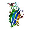
| ||||||||
|---|---|---|---|---|---|---|---|---|---|
| 1 |
| ||||||||
| Unit cell |
|
-Components
| #1: Protein | Mass: 16240.451 Da / Num. of mol.: 1 / Fragment: CALCIUM/PHOSPHOLIPID BINDING DOMAIN Source method: isolated from a genetically manipulated source Source: (gene. exp.)  References: UniProt: P05696, Transferases; Transferring phosphorus-containing groups; Phosphotransferases with an alcohol group as acceptor | ||||||
|---|---|---|---|---|---|---|---|
| #2: Chemical |   Mass: 40.078 Da / Num. of mol.: 2 / Source method: obtained synthetically / Formula: Ca Mass: 40.078 Da / Num. of mol.: 2 / Source method: obtained synthetically / Formula: Ca#3: Chemical | ChemComp-PO4 / |   Mass: 94.971 Da / Num. of mol.: 1 / Source method: obtained synthetically / Formula: PO4 Mass: 94.971 Da / Num. of mol.: 1 / Source method: obtained synthetically / Formula: PO4#4: Chemical | ChemComp-PSF / |   Mass: 455.437 Da / Num. of mol.: 1 / Source method: obtained synthetically / Formula: C18H34NO10P Mass: 455.437 Da / Num. of mol.: 1 / Source method: obtained synthetically / Formula: C18H34NO10P#5: Water | ChemComp-HOH / |  Mass: 18.015 Da / Num. of mol.: 39 / Source method: isolated from a natural source / Formula: H2O Mass: 18.015 Da / Num. of mol.: 39 / Source method: isolated from a natural source / Formula: H2O |
-Experimental details
-Experiment
| Experiment | Method: X-RAY DIFFRACTION / Number of used crystals: 1 |
|---|
- Sample preparation
| Crystal | Density Matthews: 2.82 Å3/Da / Density % sol: 56.32 % | ||||||||||||||||||||||||||||||||||||||||||
|---|---|---|---|---|---|---|---|---|---|---|---|---|---|---|---|---|---|---|---|---|---|---|---|---|---|---|---|---|---|---|---|---|---|---|---|---|---|---|---|---|---|---|---|
| Crystal grow | Temperature: 293 K / Method: vapor diffusion, hanging drop / pH: 6.5 Details: PEG8K, POTASSIUM PHOSPHATE, pH 6.5, VAPOR DIFFUSION, HANGING DROP, temperature 293K | ||||||||||||||||||||||||||||||||||||||||||
| Crystal | *PLUS Density % sol: 50 % | ||||||||||||||||||||||||||||||||||||||||||
| Crystal grow | *PLUS Temperature: 20 ℃ | ||||||||||||||||||||||||||||||||||||||||||
| Components of the solutions | *PLUS
|
-Data collection
| Diffraction | Mean temperature: 293 K |
|---|---|
| Diffraction source | Source: ROTATING ANODE / Type: RIGAKU / Wavelength: 1.5418 |
| Detector | Type: MARRESEARCH / Detector: IMAGE PLATE / Date: Feb 25, 1999 |
| Radiation | Protocol: SINGLE WAVELENGTH / Monochromatic (M) / Laue (L): M / Scattering type: x-ray |
| Radiation wavelength | Wavelength: 1.5418 Å / Relative weight: 1 |
| Reflection | Resolution: 2.6→20 Å / Num. all: 5950 / Num. obs: 5610 / % possible obs: 94.3 % / Observed criterion σ(F): 0 / Observed criterion σ(I): 0 / Redundancy: 2.9 % / Biso Wilson estimate: 36.9 Å2 / Rmerge(I) obs: 0.092 / Net I/σ(I): 9.4 |
| Reflection shell | Resolution: 2.6→2.69 Å / Redundancy: 3.9 % / Rmerge(I) obs: 0.281 / Num. unique all: 562 / % possible all: 95.1 |
| Reflection | *PLUS Num. measured all: 17072 |
| Reflection shell | *PLUS % possible obs: 95.1 % |
- Processing
| Software |
| ||||||||||||||||||||
|---|---|---|---|---|---|---|---|---|---|---|---|---|---|---|---|---|---|---|---|---|---|
| Refinement | Resolution: 2.6→18 Å / σ(F): 2 / Stereochemistry target values: Engh & Huber
| ||||||||||||||||||||
| Refinement step | Cycle: LAST / Resolution: 2.6→18 Å
| ||||||||||||||||||||
| Refine LS restraints |
| ||||||||||||||||||||
| Software | *PLUS Name: 'CNS' / Classification: refinement | ||||||||||||||||||||
| Refinement | *PLUS Rfactor Rfree: 0.233 / Rfactor Rwork: 0.195 | ||||||||||||||||||||
| Solvent computation | *PLUS | ||||||||||||||||||||
| Displacement parameters | *PLUS | ||||||||||||||||||||
| Refine LS restraints | *PLUS
|
