 Movie
Movie Controller

[English] 日本語
 Yorodumi
- PDB-2dp3: Crystal structure of a double mutant (C202A/A198V) of Triosephosp... -
+ Open data

- Basic information
| Entry | Database: PDB / ID: 2dp3 | ||||||
|---|---|---|---|---|---|---|---|
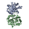
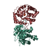
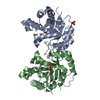
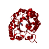
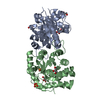
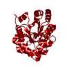
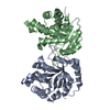
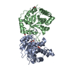
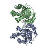
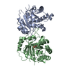
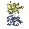
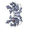
| Title | Crystal structure of a double mutant (C202A/A198V) of Triosephosphate isomerase from giardia lamblia | ||||||
 Components Components | Triosephosphate isomerase | ||||||
 Keywords Keywords | ISOMERASE / TRIOSEPHOSPHATE ISOMERASE / GIARDIA / ENZYME / ALPHA/BETA BARREL | ||||||
| Function / homology |  Function and homology information Function and homology informationmethylglyoxal synthase / methylglyoxal synthase activity / triose-phosphate isomerase / triose-phosphate isomerase activity / glyceraldehyde-3-phosphate biosynthetic process / glycerol catabolic process / glycolytic process / gluconeogenesis / cytosol Similarity search - Function | ||||||
| Biological species |  Giardia intestinalis (eukaryote) Giardia intestinalis (eukaryote) | ||||||
| Method |  X-RAY DIFFRACTION / MOLECULAR REPLACEMENT / Resolution: 2.1 Å X-RAY DIFFRACTION / MOLECULAR REPLACEMENT / Resolution: 2.1 Å | ||||||
 Authors Authors | Diaz, A. / Reyes-Vivas, H. / Lopez-Velazquez, G. | ||||||
 Citation Citation | Journal: J.Mol.Biol. / Year: 2007 Title: Disulfide Bridges in the Mesophilic Triosephosphate Isomerase from Giardia lamblia Are Related to Oligomerization and Activity Authors: Reyes-Vivas, H. / Diaz, A. / Peon, J. / Mendoza-Hernandez, G. / Hernandez-Alcantara, G. / De la Mora-De la Mora, I. / Enriquez-Flores, S. / Dominguez-Ramirez, L. / Lopez-Velazquez, G. #1: Journal: Proteins / Year: 2004 Title: An unusual triosephosphate isomerase from the early divergent eukaryote Giardia lamblia Authors: Lopez-Velazquez, G. / Molina-Ortiz, D. / Cabrera, N. / Peon-Peralta, J. / Reyes-Vivas, H. | ||||||
| History |
|
- Structure visualization
| Structure viewer | Molecule: MolmilJmol/JSmol |
|---|
- Downloads & links
-Download
| PDBx/mmCIF format | 2dp3.cif.gz | 69.5 KB | Display | PDBx/mmCIF format |
|---|---|---|---|---|
| PDB format | pdb2dp3.ent.gz | 50.4 KB | Display | PDB format |
| PDBx/mmJSON format | 2dp3.json.gz | Tree view | PDBx/mmJSON format | |
| Others |  Other downloads Other downloads |
-Validation report
| Summary document | 2dp3_validation.pdf.gz | 440.7 KB | Display | wwPDB validaton report |
|---|---|---|---|---|
| Full document | 2dp3_full_validation.pdf.gz | 443.3 KB | Display | |
| Data in XML | 2dp3_validation.xml.gz | 15 KB | Display | |
| Data in CIF | 2dp3_validation.cif.gz | 22.3 KB | Display | |
| Arichive directory | https://data.pdbj.org/pub/pdb/validation_reports/dp/2dp3ftp://data.pdbj.org/pub/pdb/validation_reports/dp/2dp3 | HTTPS FTP |
-Related structure data


-Links
 PDBj
PDBj
- Assembly
| Deposited unit | 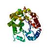
| ||||||||
|---|---|---|---|---|---|---|---|---|---|
| 1 | 
| ||||||||
| Unit cell |
| ||||||||
| Details | The second part of the biological assembly is generated by the two fold axis: |
-Components
| #1: Protein | Mass: 27936.176 Da / Num. of mol.: 1 / Mutation: C202A Source method: isolated from a genetically manipulated source Source: (gene. exp.) Giardia intestinalis (eukaryote) / Strain: WB STRAIN / Gene: gltim / Plasmid: PET3A / Production host:  | ||
|---|---|---|---|
| #2: Chemical | ChemComp-SO4 /   Mass: 96.063 Da / Num. of mol.: 4 / Source method: obtained synthetically / Formula: SO4 Mass: 96.063 Da / Num. of mol.: 4 / Source method: obtained synthetically / Formula: SO4#3: Water | ChemComp-HOH / |  Mass: 18.015 Da / Num. of mol.: 303 / Source method: isolated from a natural source / Formula: H2O Mass: 18.015 Da / Num. of mol.: 303 / Source method: isolated from a natural source / Formula: H2O |
-Experimental details
-Experiment
| Experiment | Method: X-RAY DIFFRACTION / Number of used crystals: 1 |
|---|
- Sample preparation
| Crystal | Density Matthews: 2.96 Å3/Da / Density % sol: 58.38 % |
|---|---|
| Crystal grow | Temperature: 291 K / Method: vapor diffusion, sitting drop / pH: 7.4 Details: 5microL of C202A (13mg/mL) dissolved in 100mM triethanolamine, 10mM EDTA, mixed with 5microL of 2M ammonium sulfate,5% isopropanol, pH 7.4, VAPOR DIFFUSION, SITTING DROP, temperature 291K |
-Data collection
| Diffraction | Mean temperature: 113 K |
|---|---|
| Diffraction source | Source: ROTATING ANODE / Type: RIGAKU RU200 / Wavelength: 1.5418 Å |
| Detector | Type: RIGAKU RAXIS IIC / Detector: IMAGE PLATE / Date: Jun 11, 2004 / Details: mirrors |
| Radiation | Monochromator: YALE MIRRORS / Protocol: SINGLE WAVELENGTH / Monochromatic (M) / Laue (L): M / Scattering type: x-ray |
| Radiation wavelength | Wavelength: 1.5418 Å / Relative weight: 1 |
| Reflection | Resolution: 2.1→28.65 Å / Num. obs: 18609 / % possible obs: 94.1 % / Observed criterion σ(F): 0 / Observed criterion σ(I): 0 / Redundancy: 2.1 % / Biso Wilson estimate: 14.2 Å2 / Rmerge(I) obs: 0.071 |
| Reflection shell | Resolution: 2.1→2.23 Å / Redundancy: 2 % / Rmerge(I) obs: 0.065 / Mean I/σ(I) obs: 8.1 / Num. unique all: 2875 / % possible all: 98.6 |
- Processing
| Software |
| ||||||||||||||||||||
|---|---|---|---|---|---|---|---|---|---|---|---|---|---|---|---|---|---|---|---|---|---|
| Refinement | Method to determine structure: MOLECULAR REPLACEMENT Starting model: PDB entry 1tcd, 1lyx, 1m6j Resolution: 2.1→28.65 Å / Cross valid method: THROUGHOUT / σ(F): 0 / σ(I): 0 / Stereochemistry target values: Engh & Huber
| ||||||||||||||||||||
| Displacement parameters |
| ||||||||||||||||||||
| Refine analyze | Luzzati coordinate error obs: 0.2 Å / Luzzati sigma a obs: 0.09 Å | ||||||||||||||||||||
| Refinement step | Cycle: LAST / Resolution: 2.1→28.65 Å
| ||||||||||||||||||||
| Refine LS restraints |
| ||||||||||||||||||||
| LS refinement shell | Resolution: 2.1→2.23 Å / Rfactor Rfree error: 0.009
|
