Mass: 18.015 Da / Num. of mol.: 230 / Source method: isolated from a natural source / Formula: H2O
Compound details
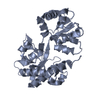
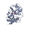
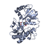
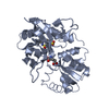
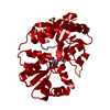
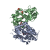
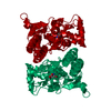
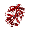
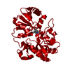
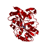
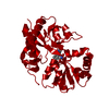
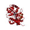
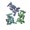
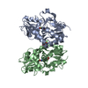
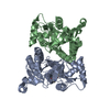
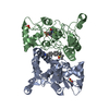
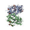
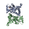
L-GLUTAMATE ACTS AS AN EXCITATORY NEUROTRANSMITTER AT MANY SYNAPSES IN THE CENTRAL NERVOUS SYSTEM
Has protein modification
Y
Sequence details
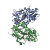
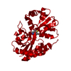
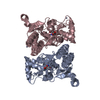
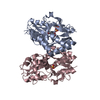
NATIVE GLUR2 IS A MEMBRANE PROTEIN. THE PROTEIN CRYSTALLIZED IS THE EXTRACELLULAR LIGAND-BINDING ...NATIVE GLUR2 IS A MEMBRANE PROTEIN. THE PROTEIN CRYSTALLIZED IS THE EXTRACELLULAR LIGAND-BINDING CORE OF GLUR2. TRANSMEMBRANE REGIONS WERE GENETICALLY REMOVED AND REPLACED WITH A GLY-THR LINKER (RESIDUES 115-116). THEREFORE, THE SEQUENCE MATCHES DISCONTINUOUSLY WITH THE REFERENCE DATABASE (413-527, 653-796). THE TWO FIRST RESIDUES OF THE SEQUENCE (GLY, ALA) ARE CLONING ARTIFACTS AND WERE NOT LOCATED IN THE ELECTRON DENSITY MAP.
-
Experimental details
-
Experiment
Experiment
Method: X-RAY DIFFRACTION / Number of used crystals: 1
-
Sample preparation
Crystal
Density Matthews: 2.4 Å3/Da / Density % sol: 48.73 %
In the structure databanks used in Yorodumi, some data are registered as the other names, "COVID-19 virus" and "2019-nCoV". Here are the details of the virus and the list of structure data.
Jan 31, 2019. EMDB accession codes are about to change! (news from PDBe EMDB page)
EMDB accession codes are about to change! (news from PDBe EMDB page)
The allocation of 4 digits for EMDB accession codes will soon come to an end. Whilst these codes will remain in use, new EMDB accession codes will include an additional digit and will expand incrementally as the available range of codes is exhausted. The current 4-digit format prefixed with “EMD-” (i.e. EMD-XXXX) will advance to a 5-digit format (i.e. EMD-XXXXX), and so on. It is currently estimated that the 4-digit codes will be depleted around Spring 2019, at which point the 5-digit format will come into force.
The EM Navigator/Yorodumi systems omit the EMD- prefix.
Related info.:Q: What is EMD? / ID/Accession-code notation in Yorodumi/EM Navigator
Yorodumi is a browser for structure data from EMDB, PDB, SASBDB, etc.
This page is also the successor to EM Navigator detail page, and also detail information page/front-end page for Omokage search.
The word "yorodu" (or yorozu) is an old Japanese word meaning "ten thousand". "mi" (miru) is to see.
Related info.:EMDB / PDB / SASBDB / Comparison of 3 databanks / Yorodumi Search / Aug 31, 2016. New EM Navigator & Yorodumi / Yorodumi Papers / Jmol/JSmol / Function and homology information / Changes in new EM Navigator and Yorodumi
 Movie
Movie Controller

 Yorodumi
 Open data
 Basic information
 Components
 Keywords
 Function and homology information
 X-RAY DIFFRACTION /
  Authors
 Citation
 Structure visualization
 Downloads & links
 Other downloads


 PDBj
PDBj


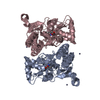
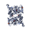
 Assembly


 Mass: 96.063 Da / Num. of mol.: 4 / Source method: obtained synthetically / Formula: SO4
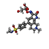
 Mass: 518.583 Da / Num. of mol.: 1 / Source method: obtained synthetically / Formula: C24H30N4O7S
 Type: L-peptide linking / Mass: 147.129 Da / Num. of mol.: 1 / Source method: obtained synthetically / Formula: C5H9NO4
 Mass: 18.015 Da / Num. of mol.: 230 / Source method: isolated from a natural source / Formula: H2O
 Sample preparation
 Processing