 Movie
Movie Controller

[English] 日本語
 Yorodumi
- PDB-2aix: X-ray structure of the GLUR2 ligand-binding core (S1S2J) in compl... -
+ Open data

- Basic information
| Entry | Database: PDB / ID: 2aix | ||||||
|---|---|---|---|---|---|---|---|
| Title | X-ray structure of the GLUR2 ligand-binding core (S1S2J) in complex with (s)-thio-atpa at 2.2 a resolution. | ||||||
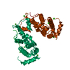
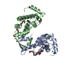
 Components Components | Glutamate receptor 2 | ||||||
 Keywords Keywords | MEMBRANE PROTEIN / IONOTROPIC GLUTAMATE RECEPTOR GLUR2 / LIGAND-BINDING CORE / AGONIST COMPLEX. | ||||||
| Function / homology |  Function and homology information Function and homology informationspine synapse / dendritic spine neck / dendritic spine cytoplasm / dendritic spine head / cellular response to amine stimulus / Activation of AMPA receptors / ligand-gated monoatomic cation channel activity / perisynaptic space / Trafficking of GluR2-containing AMPA receptors / response to lithium ion ...spine synapse / dendritic spine neck / dendritic spine cytoplasm / dendritic spine head / cellular response to amine stimulus / Activation of AMPA receptors / ligand-gated monoatomic cation channel activity / perisynaptic space / Trafficking of GluR2-containing AMPA receptors / response to lithium ion / AMPA glutamate receptor activity / AMPA glutamate receptor clustering / kainate selective glutamate receptor activity / immunoglobulin binding / AMPA glutamate receptor complex / regulation of receptor recycling / extracellularly glutamate-gated ion channel activity / cellular response to glycine / ionotropic glutamate receptor complex / asymmetric synapse / Unblocking of NMDA receptors, glutamate binding and activation / glutamate receptor binding / positive regulation of synaptic transmission / conditioned place preference / response to fungicide / regulation of synaptic transmission, glutamatergic / extracellular ligand-gated monoatomic ion channel activity / cytoskeletal protein binding / glutamate-gated receptor activity / cellular response to brain-derived neurotrophic factor stimulus / regulation of long-term synaptic depression / somatodendritic compartment / glutamate-gated calcium ion channel activity / presynaptic active zone membrane / excitatory synapse / ionotropic glutamate receptor signaling pathway / ionotropic glutamate receptor binding / dendrite cytoplasm / dendrite membrane / ligand-gated monoatomic ion channel activity involved in regulation of presynaptic membrane potential / positive regulation of excitatory postsynaptic potential / dendritic shaft / SNARE binding / synaptic membrane / PDZ domain binding / establishment of protein localization / protein tetramerization / synaptic transmission, glutamatergic / transmitter-gated monoatomic ion channel activity involved in regulation of postsynaptic membrane potential / cerebral cortex development / receptor internalization / postsynaptic density membrane / modulation of chemical synaptic transmission / Schaffer collateral - CA1 synapse / terminal bouton / synaptic vesicle / long-term synaptic potentiation / amyloid-beta binding / synaptic vesicle membrane / growth cone / presynapse / signaling receptor activity / presynaptic membrane / scaffold protein binding / dendritic spine / chemical synaptic transmission / perikaryon / postsynaptic membrane / neuron projection / postsynaptic density / external side of plasma membrane / axon / neuronal cell body / synapse / dendrite / protein kinase binding / protein-containing complex binding / glutamatergic synapse / cell surface / endoplasmic reticulum / protein-containing complex / membrane / identical protein binding / plasma membrane Similarity search - Function | ||||||
| Biological species |  | ||||||
| Method |  X-RAY DIFFRACTION / SYNCHROTRON / MOLECULAR REPLACEMENT / Resolution: 2.17 Å X-RAY DIFFRACTION / SYNCHROTRON / MOLECULAR REPLACEMENT / Resolution: 2.17 Å | ||||||
 Authors Authors | Kastrup, J.S. | ||||||
 Citation Citation | Journal: To be Published Title: Distinct kinetics of ionotropic glutamate receptor 2 splice variants characterized by fast agonist application and crystallization of a receptor-ligand complex. Authors: Holm, M.M. / Lunn, M.L. / Traynelis, S.F. / Kastrup, J.S. / Egebjerg, J. #1: Journal: J.Mol.Biol. / Year: 2002Title: Structural Basis for Ampa Receptor Activation and Ligand Selectivity: Crystal Structures of Five Agonist Complexes with the Glur2 Ligand Binding Core. Authors: Hogner, A. / Kastrup, J.S. / Jin, R. / Liljefors, T. / Mayer, M.L. / Egebjerg, J. / Larsen, I. / Gouaux, E. #2: Journal: Neuron / Year: 2000Title: Mechanisms for Activation and Antagonism of an Ampa-Sensitive Glutamate Receptor: Crystal Structures of the Glur2 Ligand Binding Core. Authors: Armstrong, N. / Gouaux, E. #3: Journal: Nature / Year: 2002Title: Mechanism of Glutamate Receptor Desensitization. Authors: Sun, Y. / Olson, R. / Horning, M. / Armstrong, N. / Mayer, M. / Gouaux, E. #4: Journal: Protein Sci. / Year: 1998Title: Probing the Ligand Binding Domain of the Glur2 Receptor by Proteolysis and Deletion Mutagenesis Defines Domain Boundaries and Yields a Crystallizable Construct. Authors: Chen, G.Q. / Sun, R. / Jin, R. / Gouaux, E. | ||||||
| History |
| ||||||
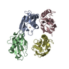
| Remark 300 | BIOMOLECULE: 1 THIS ENTRY CONTAINS THE CRYSTALLOGRAPHIC ASYMMETRIC UNIT WHICH CONSISTS OF 1 CHAIN(S) ...BIOMOLECULE: 1 THIS ENTRY CONTAINS THE CRYSTALLOGRAPHIC ASYMMETRIC UNIT WHICH CONSISTS OF 1 CHAIN(S). THE RECEPTOR CONSISTS OF FOUR SUBUNITS CONSTITUTING A TETRAMER. ONE DIMER OF THE TETRAMER CAN BE GENERATED IN THE CRYSTAL. SEE REMARK 350 FOR INFORMATION ON GENERATING THE BIOLOGICAL MOLECULE(S). | ||||||
| Remark 999 | SEQUENCE Native GluR2 is a membrane protein. The protein crystallized is the extracellular ligand- ...SEQUENCE Native GluR2 is a membrane protein. The protein crystallized is the extracellular ligand-binding core of GluR2. Transmembrane regions were genetically removed and replaced with a Gly-Thr linker (residues 115-116). Therefore, the sequence matches discontinuously with the reference database (413-527, 653-796). |
- Structure visualization
| Structure viewer | Molecule: MolmilJmol/JSmol |
|---|
- Downloads & links
-Download
| PDBx/mmCIF format | 2aix.cif.gz | 70.8 KB | Display | PDBx/mmCIF format |
|---|---|---|---|---|
| PDB format | pdb2aix.ent.gz | 50.9 KB | Display | PDB format |
| PDBx/mmJSON format | 2aix.json.gz | Tree view | PDBx/mmJSON format | |
| Others |  Other downloads Other downloads |
-Validation report
| Arichive directory | https://data.pdbj.org/pub/pdb/validation_reports/ai/2aixftp://data.pdbj.org/pub/pdb/validation_reports/ai/2aix | HTTPS FTP |
|---|
-Related structure data
| Related structure data |  1m5bS S: Starting model for refinement |
|---|---|
| Similar structure data |


-Links
 PDBj
PDBj


- Assembly
| Deposited unit | 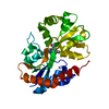
| ||||||||
|---|---|---|---|---|---|---|---|---|---|
| 1 | 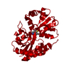
| ||||||||
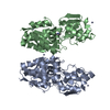
| Unit cell |
|
-Components
| #1: Protein | Mass: 29221.682 Da / Num. of mol.: 1 / Fragment: GLUR2-FLOP LIGAND-BINDING CORE (S1S2J). Source method: isolated from a genetically manipulated source Source: (gene. exp.)  |
|---|---|
| #2: Chemical | ChemComp-U1K / (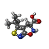  Mass: 244.311 Da / Num. of mol.: 1 / Source method: obtained synthetically / Formula: C10H16N2O3S Mass: 244.311 Da / Num. of mol.: 1 / Source method: obtained synthetically / Formula: C10H16N2O3S |
| #3: Water | ChemComp-HOH /  Mass: 18.015 Da / Num. of mol.: 234 / Source method: isolated from a natural source / Formula: H2O Mass: 18.015 Da / Num. of mol.: 234 / Source method: isolated from a natural source / Formula: H2O |
| Has protein modification | Y |
-Experimental details
-Experiment
| Experiment | Method: X-RAY DIFFRACTION / Number of used crystals: 1 |
|---|
- Sample preparation
| Crystal | Density Matthews: 2.42 Å3/Da / Density % sol: 48.8 % |
|---|---|
| Crystal grow | Temperature: 279 K / pH: 6.5 Details: SODIUM CHLORIDE, CACODYLATE, PEG 8000, pH 6.50, VAPOR DIFFUSION, HANGING DROP, temperature 279K |
-Data collection
| Diffraction | Mean temperature: 100 K |
|---|---|
| Diffraction source | Source: SYNCHROTRON / Site: MAX II  / Beamline: I711 / Wavelength: 1.0835 / Beamline: I711 / Wavelength: 1.0835 |
| Detector | Type: MARRESEARCH / Detector: IMAGE PLATE / Date: Oct 14, 2000 |
| Radiation | Protocol: SINGLE WAVELENGTH / Monochromatic (M) / Laue (L): M / Scattering type: x-ray |
| Radiation wavelength | Wavelength: 1.0835 Å / Relative weight: 1 |
| Reflection | Resolution: 2.17→20 Å / Num. obs: 15624 / % possible obs: 98.3 % / Observed criterion σ(I): 0 / Redundancy: 6.3 % / Biso Wilson estimate: 24.5 Å2 / Rmerge(I) obs: 0.067 / Net I/σ(I): 24.6 |
| Reflection shell | Resolution: 2.17→2.25 Å / Rmerge(I) obs: 0.228 / Mean I/σ(I) obs: 7.6 / % possible all: 93.4 |
- Processing
| Software |
| ||||||||||||||||||||||||||||||||||||||||||||||||||||||||||||||||||||||||||||||||
|---|---|---|---|---|---|---|---|---|---|---|---|---|---|---|---|---|---|---|---|---|---|---|---|---|---|---|---|---|---|---|---|---|---|---|---|---|---|---|---|---|---|---|---|---|---|---|---|---|---|---|---|---|---|---|---|---|---|---|---|---|---|---|---|---|---|---|---|---|---|---|---|---|---|---|---|---|---|---|---|---|---|
| Refinement | Method to determine structure: MOLECULAR REPLACEMENT Starting model: PDB ENTRY 1M5B. Resolution: 2.17→20 Å / Rfactor Rfree error: 0.008 / Data cutoff high absF: 1987987.79 / Data cutoff low absF: 0 / Isotropic thermal model: RESTRAINED. / Cross valid method: THROUGHOUT / σ(F): 0 / Stereochemistry target values: ENGH & HUBER Details: THE FIRST THREE N-TERMINAL RESIDUES AND THE LAST TWO C-TERMINAL RESIDUES WERE NOT LOCATED IN THE ELECTRON DENSITY MAP.
| ||||||||||||||||||||||||||||||||||||||||||||||||||||||||||||||||||||||||||||||||
| Solvent computation | Solvent model: FLAT MODEL / Bsol: 60.3512 Å2 / ksol: 0.414103 e/Å3 | ||||||||||||||||||||||||||||||||||||||||||||||||||||||||||||||||||||||||||||||||
| Displacement parameters | Biso mean: 25.7 Å2
| ||||||||||||||||||||||||||||||||||||||||||||||||||||||||||||||||||||||||||||||||
| Refine analyze |
| ||||||||||||||||||||||||||||||||||||||||||||||||||||||||||||||||||||||||||||||||
| Refinement step | Cycle: LAST / Resolution: 2.17→20 Å
| ||||||||||||||||||||||||||||||||||||||||||||||||||||||||||||||||||||||||||||||||
| Refine LS restraints |
| ||||||||||||||||||||||||||||||||||||||||||||||||||||||||||||||||||||||||||||||||
| LS refinement shell | Resolution: 2.17→2.31 Å / Rfactor Rfree error: 0.023 / Total num. of bins used: 6
| ||||||||||||||||||||||||||||||||||||||||||||||||||||||||||||||||||||||||||||||||
| Xplor file |
|
