 Movie
Movie Controller

[English] 日本語
 Yorodumi
- PDB-1wvj: Exploring the GluR2 ligand-binding core in complex with the bicyc... -
+ Open data

- Basic information
| Entry | Database: PDB / ID: 1wvj | ||||||
|---|---|---|---|---|---|---|---|
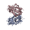
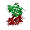
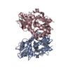
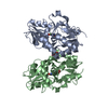
| Title | Exploring the GluR2 ligand-binding core in complex with the bicyclic AMPA analogue (S)-4-AHCP | ||||||
 Components Components | ionotropic glutamate receptor 2 | ||||||
 Keywords Keywords | MEMBRANE PROTEIN / Ionotropic glutamate receptor GluR2 / ligand-binding core / complex with bicyclic AMPA analogue | ||||||
| Function / homology |  Function and homology information Function and homology informationspine synapse / dendritic spine neck / dendritic spine cytoplasm / dendritic spine head / cellular response to amine stimulus / Activation of AMPA receptors / ligand-gated monoatomic cation channel activity / perisynaptic space / Trafficking of GluR2-containing AMPA receptors / response to lithium ion ...spine synapse / dendritic spine neck / dendritic spine cytoplasm / dendritic spine head / cellular response to amine stimulus / Activation of AMPA receptors / ligand-gated monoatomic cation channel activity / perisynaptic space / Trafficking of GluR2-containing AMPA receptors / response to lithium ion / AMPA glutamate receptor activity / AMPA glutamate receptor clustering / kainate selective glutamate receptor activity / immunoglobulin binding / AMPA glutamate receptor complex / regulation of receptor recycling / extracellularly glutamate-gated ion channel activity / cellular response to glycine / ionotropic glutamate receptor complex / asymmetric synapse / Unblocking of NMDA receptors, glutamate binding and activation / glutamate receptor binding / positive regulation of synaptic transmission / conditioned place preference / response to fungicide / regulation of synaptic transmission, glutamatergic / extracellular ligand-gated monoatomic ion channel activity / cytoskeletal protein binding / glutamate-gated receptor activity / cellular response to brain-derived neurotrophic factor stimulus / regulation of long-term synaptic depression / somatodendritic compartment / glutamate-gated calcium ion channel activity / presynaptic active zone membrane / excitatory synapse / ionotropic glutamate receptor signaling pathway / ionotropic glutamate receptor binding / dendrite cytoplasm / dendrite membrane / ligand-gated monoatomic ion channel activity involved in regulation of presynaptic membrane potential / positive regulation of excitatory postsynaptic potential / dendritic shaft / SNARE binding / synaptic membrane / PDZ domain binding / establishment of protein localization / protein tetramerization / synaptic transmission, glutamatergic / transmitter-gated monoatomic ion channel activity involved in regulation of postsynaptic membrane potential / cerebral cortex development / receptor internalization / postsynaptic density membrane / modulation of chemical synaptic transmission / Schaffer collateral - CA1 synapse / terminal bouton / synaptic vesicle / long-term synaptic potentiation / amyloid-beta binding / synaptic vesicle membrane / growth cone / presynapse / signaling receptor activity / presynaptic membrane / scaffold protein binding / dendritic spine / chemical synaptic transmission / perikaryon / postsynaptic membrane / neuron projection / postsynaptic density / external side of plasma membrane / axon / neuronal cell body / synapse / dendrite / protein kinase binding / protein-containing complex binding / glutamatergic synapse / cell surface / endoplasmic reticulum / protein-containing complex / membrane / identical protein binding / plasma membrane Similarity search - Function | ||||||
| Biological species |  | ||||||
| Method |  X-RAY DIFFRACTION / SYNCHROTRON / MOLECULAR REPLACEMENT / Resolution: 1.75 Å X-RAY DIFFRACTION / SYNCHROTRON / MOLECULAR REPLACEMENT / Resolution: 1.75 Å | ||||||
 Authors Authors | Nielsen, B.B. / Pickering, D.S. / Greenwood, J.R. / Brehm, L. / Gajhede, M. / Schousboe, A. / Kastrup, J.S. | ||||||
 Citation Citation | Journal: FEBS J. / Year: 2005 Title: Exploring the GluR2 ligand-binding core in complex with the bicyclical AMPA analogue (S)-4-AHCP Authors: Nielsen, B.B. / Pickering, D.S. / Greenwood, J.R. / Brehm, L. / Gajhede, M. / Schousboe, A. / Kastrup, J.S. #1: Journal: J.Mol.Biol. / Year: 2002Title: Structural basis for AMPA receptor activation and ligand selectivity: crystal structures of five agonist complexes with the GluR2 ligand-binding core Authors: Hogner, A. / Kastrup, J.S. / Jin, R. / Liljefors, T. / Mayer, M.L. / Egebjerg, J. / Larsen, I.K. / Gouaux, E. #2: Journal: Neuron / Year: 2000Title: Mechanisms for activation and antagonism of an AMPA-sensitive glutamate receptor: crystal structures of the GluR2 ligand binding core Authors: Armstrong, N. / Gouaux, E. #3: Journal: Protein Sci. / Year: 1998 Title: Probing the ligand binding domain of the GluR2 receptor by proteolysis and deletion mutagenesis defines domain boundaries and yields a crystallizable construct Authors: Chen, G.Q. / Sun, Y. / Jin, R. / Gouaux, E. | ||||||
| History |
|
- Structure visualization
| Structure viewer | Molecule: MolmilJmol/JSmol |
|---|
- Downloads & links
-Download
| PDBx/mmCIF format | 1wvj.cif.gz | 74.3 KB | Display | PDBx/mmCIF format |
|---|---|---|---|---|
| PDB format | pdb1wvj.ent.gz | 54.5 KB | Display | PDB format |
| PDBx/mmJSON format | 1wvj.json.gz | Tree view | PDBx/mmJSON format | |
| Others |  Other downloads Other downloads |
-Validation report
| Arichive directory | https://data.pdbj.org/pub/pdb/validation_reports/wv/1wvjftp://data.pdbj.org/pub/pdb/validation_reports/wv/1wvj | HTTPS FTP |
|---|
-Related structure data
| Related structure data |  1m5cS S: Starting model for refinement |
|---|---|
| Similar structure data |


-Links
 PDBj
PDBj


- Assembly
| Deposited unit | 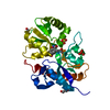
| ||||||||
|---|---|---|---|---|---|---|---|---|---|
| 1 | 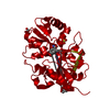
| ||||||||
| Unit cell |
| ||||||||
| Components on special symmetry positions |
|
-Components
| #1: Protein | Mass: 29221.682 Da / Num. of mol.: 1 Source method: isolated from a genetically manipulated source Details: Flop ligand-binding core of GluR2(GluR2-S1S2J) / Source: (gene. exp.)  | ||||||||
|---|---|---|---|---|---|---|---|---|---|
| #2: Chemical |   Mass: 96.063 Da / Num. of mol.: 2 / Source method: obtained synthetically / Formula: SO4 Mass: 96.063 Da / Num. of mol.: 2 / Source method: obtained synthetically / Formula: SO4#3: Chemical | ChemComp-IBC / |   Mass: 238.240 Da / Num. of mol.: 1 / Source method: obtained synthetically / Formula: C11H14N2O4 Mass: 238.240 Da / Num. of mol.: 1 / Source method: obtained synthetically / Formula: C11H14N2O4#4: Chemical |   Mass: 92.094 Da / Num. of mol.: 3 / Source method: obtained synthetically / Formula: C3H8O3 Mass: 92.094 Da / Num. of mol.: 3 / Source method: obtained synthetically / Formula: C3H8O3#5: Water | ChemComp-HOH / |  Mass: 18.015 Da / Num. of mol.: 309 / Source method: isolated from a natural source / Formula: H2O Mass: 18.015 Da / Num. of mol.: 309 / Source method: isolated from a natural source / Formula: H2OHas protein modification | Y | |
-Experimental details
-Experiment
| Experiment | Method: X-RAY DIFFRACTION / Number of used crystals: 1 |
|---|
- Sample preparation
| Crystal | Density Matthews: 2.3 Å3/Da / Density % sol: 46.1 % |
|---|---|
| Crystal grow | Temperature: 279 K / Method: vapor diffusion, hanging drop / pH: 4.5 Details: PEG 3350, phosphate-citrate buffer, lithium sulfate, pH 4.5, VAPOR DIFFUSION, HANGING DROP, temperature 279K |
-Data collection
| Diffraction | Mean temperature: 110 K |
|---|---|
| Diffraction source | Source: SYNCHROTRON / Site: EMBL/DESY, HAMBURG  / Beamline: X11 / Wavelength: 0.811 Å / Beamline: X11 / Wavelength: 0.811 Å |
| Detector | Type: MARRESEARCH / Detector: CCD / Date: Nov 25, 2003 |
| Radiation | Protocol: SINGLE WAVELENGTH / Monochromatic (M) / Laue (L): M / Scattering type: x-ray |
| Radiation wavelength | Wavelength: 0.811 Å / Relative weight: 1 |
| Reflection | Resolution: 1.75→20.08 Å / Num. all: 27472 / Num. obs: 27472 / % possible obs: 99.1 % / Observed criterion σ(F): -3 / Observed criterion σ(I): -3 / Redundancy: 3.5 % / Biso Wilson estimate: 18.1 Å2 / Rsym value: 0.037 / Net I/σ(I): 26.7 |
| Reflection shell | Resolution: 1.75→1.78 Å / Mean I/σ(I) obs: 4.1 / Num. unique all: 1333 / Rsym value: 0.227 / % possible all: 96.3 |
- Processing
| Software |
| ||||||||||||||||||||||||||||||||||||||||||||||||||||||||||||||||||||||
|---|---|---|---|---|---|---|---|---|---|---|---|---|---|---|---|---|---|---|---|---|---|---|---|---|---|---|---|---|---|---|---|---|---|---|---|---|---|---|---|---|---|---|---|---|---|---|---|---|---|---|---|---|---|---|---|---|---|---|---|---|---|---|---|---|---|---|---|---|---|---|---|
| Refinement | Method to determine structure: MOLECULAR REPLACEMENT Starting model: 1M5C Resolution: 1.75→20.08 Å / Cor.coef. Fo:Fc: 0.959 / Cor.coef. Fo:Fc free: 0.936 / SU B: 2.37 / SU ML: 0.077 / Cross valid method: THROUGHOUT / σ(F): 0 / ESU R: 0.116 / ESU R Free: 0.117 / Stereochemistry target values: MAXIMUM LIKELIHOOD Details: The side chains of the following residues are not fully defined: LYS A4, LYS A21, GLU A24, GLU A27, LYS A50, LYS A52, ARG A64, ALA A66, ASP A67, LYS A69, LYS A82, GLU A125, LYS A129, THR ...Details: The side chains of the following residues are not fully defined: LYS A4, LYS A21, GLU A24, GLU A27, LYS A50, LYS A52, ARG A64, ALA A66, ASP A67, LYS A69, LYS A82, GLU A125, LYS A129, THR A131, GLU A145, ARG A148, ARG A149, LYS A151, LYS A157, ARG A172, GLU A176, LYS A183, LYS A204, LYS A240 and GLU A243.
| ||||||||||||||||||||||||||||||||||||||||||||||||||||||||||||||||||||||
| Solvent computation | Ion probe radii: 0.8 Å / Shrinkage radii: 0.8 Å / VDW probe radii: 1.4 Å / Solvent model: BABINET MODEL WITH MASK | ||||||||||||||||||||||||||||||||||||||||||||||||||||||||||||||||||||||
| Displacement parameters | Biso mean: 20.624 Å2
| ||||||||||||||||||||||||||||||||||||||||||||||||||||||||||||||||||||||
| Refinement step | Cycle: LAST / Resolution: 1.75→20.08 Å
| ||||||||||||||||||||||||||||||||||||||||||||||||||||||||||||||||||||||
| Refine LS restraints |
| ||||||||||||||||||||||||||||||||||||||||||||||||||||||||||||||||||||||
| LS refinement shell | Resolution: 1.754→1.8 Å / Total num. of bins used: 20
|
