 Movie
Movie Controller

[English] 日本語
 Yorodumi
- PDB-2anj: Crystal Structure of the Glur2 Ligand Binding Core (S1S2J-Y450W) ... -
+ Open data

- Basic information
| Entry | Database: PDB / ID: 2anj | ||||||
|---|---|---|---|---|---|---|---|
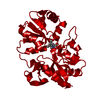
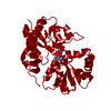
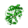
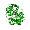
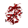
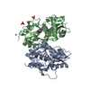
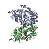
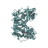
| Title | Crystal Structure of the Glur2 Ligand Binding Core (S1S2J-Y450W) Mutant in Complex With the Partial Agonist Kainic Acid at 2.1 A Resolution | ||||||
 Components Components | Glutamate receptor 2 | ||||||
 Keywords Keywords | MEMBRANE PROTEIN / Ionotropic Glutamate Receptor / GluR2 Ligand-Binding Core / Agonist Complex / Mutant | ||||||
| Function / homology |  Function and homology information Function and homology informationspine synapse / dendritic spine neck / dendritic spine cytoplasm / dendritic spine head / cellular response to amine stimulus / Activation of AMPA receptors / ligand-gated monoatomic cation channel activity / perisynaptic space / Trafficking of GluR2-containing AMPA receptors / response to lithium ion ...spine synapse / dendritic spine neck / dendritic spine cytoplasm / dendritic spine head / cellular response to amine stimulus / Activation of AMPA receptors / ligand-gated monoatomic cation channel activity / perisynaptic space / Trafficking of GluR2-containing AMPA receptors / response to lithium ion / AMPA glutamate receptor activity / AMPA glutamate receptor clustering / regulation of receptor recycling / kainate selective glutamate receptor activity / immunoglobulin binding / AMPA glutamate receptor complex / extracellularly glutamate-gated ion channel activity / cellular response to glycine / ionotropic glutamate receptor complex / asymmetric synapse / Unblocking of NMDA receptors, glutamate binding and activation / glutamate receptor binding / positive regulation of synaptic transmission / conditioned place preference / regulation of synaptic transmission, glutamatergic / response to fungicide / extracellular ligand-gated monoatomic ion channel activity / cytoskeletal protein binding / glutamate-gated receptor activity / cellular response to brain-derived neurotrophic factor stimulus / regulation of long-term synaptic depression / somatodendritic compartment / glutamate-gated calcium ion channel activity / presynaptic active zone membrane / ionotropic glutamate receptor signaling pathway / excitatory synapse / ionotropic glutamate receptor binding / dendrite cytoplasm / dendrite membrane / ligand-gated monoatomic ion channel activity involved in regulation of presynaptic membrane potential / positive regulation of excitatory postsynaptic potential / dendritic shaft / SNARE binding / synaptic membrane / PDZ domain binding / protein tetramerization / establishment of protein localization / synaptic transmission, glutamatergic / transmitter-gated monoatomic ion channel activity involved in regulation of postsynaptic membrane potential / receptor internalization / cerebral cortex development / postsynaptic density membrane / modulation of chemical synaptic transmission / Schaffer collateral - CA1 synapse / long-term synaptic potentiation / terminal bouton / synaptic vesicle / amyloid-beta binding / synaptic vesicle membrane / presynapse / growth cone / signaling receptor activity / presynaptic membrane / scaffold protein binding / chemical synaptic transmission / dendritic spine / perikaryon / postsynaptic membrane / neuron projection / postsynaptic density / external side of plasma membrane / axon / neuronal cell body / dendrite / synapse / protein kinase binding / protein-containing complex binding / glutamatergic synapse / cell surface / endoplasmic reticulum / protein-containing complex / membrane / identical protein binding / plasma membrane Similarity search - Function | ||||||
| Biological species |  | ||||||
| Method |  X-RAY DIFFRACTION / SYNCHROTRON / MOLECULAR REPLACEMENT / Resolution: 2.1 Å X-RAY DIFFRACTION / SYNCHROTRON / MOLECULAR REPLACEMENT / Resolution: 2.1 Å | ||||||
 Authors Authors | Holm, M.M. / Naur, P. / Vestergaard, B. / Geballe, M.T. / Gajhede, M. / Kastrup, J.S. / Traynelis, S.F. / Egebjerg, J. | ||||||
 Citation Citation | Journal: J.Biol.Chem. / Year: 2005 Title: A Binding Site Tyrosine Shapes Desensitization Kinetics and Agonist Potency at GluR2: a mutagenic, kinetic, and crystallographic study Authors: Holm, M.M. / Naur, P. / Vestergaard, B. / Geballe, M.T. / Gajhede, M. / Kastrup, J.S. / Traynelis, S.F. / Egebjerg, J. #1: Journal: Neuron / Year: 2000Title: Mechanisms for activation and antagonism of an AMPA-sensitive glutamate receptor: crystal structures of the GluR2 ligand binding core. Authors: Armstrong, N. / Gouaux, E. #2: Journal: Mol.Pharmacol. / Year: 2005Title: Tyr702 is an important determinant of agonist binding and domain closure of the ligand-binding core of GluR2. Authors: Frandsen, A. / Pickering, D.S. / Vestergaard, B. / Kasper, C. / Nielsen, B.B. / Greenwood, J.R. / Campiani, G. / Fattorusso, C. / Gajhede, M. / Schousboe, A. / Kastrup, J.S. #3: Journal: Proc.Natl.Acad.Sci.USA / Year: 2003Title: Tuning Activation of the Ampa-Sensitive Glur2 Ion Channel by Genetic Adjustment of Agonist-Induced Authors: Armstrong, N. / Mayer, M. / Gouaux, E. | ||||||
| History |
| ||||||
| Remark 999 | SEQUENCE Native GluR2 is a membrane protein. The protein crystallized is the extracellular ligand ...SEQUENCE Native GluR2 is a membrane protein. The protein crystallized is the extracellular ligand binding domain of GluR2. Transmembrane regions were genetically removed and replaced with a Gly-Thr linker (residues 118-119). |
- Structure visualization
| Structure viewer | Molecule: MolmilJmol/JSmol |
|---|
- Downloads & links
-Download
| PDBx/mmCIF format | 2anj.cif.gz | 71.3 KB | Display | PDBx/mmCIF format |
|---|---|---|---|---|
| PDB format | pdb2anj.ent.gz | 51.5 KB | Display | PDB format |
| PDBx/mmJSON format | 2anj.json.gz | Tree view | PDBx/mmJSON format | |
| Others |  Other downloads Other downloads |
-Validation report
| Arichive directory | https://data.pdbj.org/pub/pdb/validation_reports/an/2anjftp://data.pdbj.org/pub/pdb/validation_reports/an/2anj | HTTPS FTP |
|---|
-Related structure data
| Related structure data |  1ftkS S: Starting model for refinement |
|---|---|
| Similar structure data |


-Links
 PDBj
PDBj


- Assembly
| Deposited unit | 
| ||||||||
|---|---|---|---|---|---|---|---|---|---|
| 1 | 
| ||||||||
| Unit cell |
| ||||||||
| Details | The intact receptor is a tetramer consisting of a dimer-of-dimers. Only the dimer can be found in the crystal by applying the following transformation to chain A: -1 0 0 0 -1 0 0 0 1 1 0 0 |
-Components
| #1: Protein | Mass: 29244.721 Da / Num. of mol.: 1 / Fragment: Ligand Binding Core / Mutation: Y61W Source method: isolated from a genetically manipulated source Source: (gene. exp.)  |
|---|---|
| #2: Chemical | ChemComp-KAI /   Mass: 213.230 Da / Num. of mol.: 1 / Source method: obtained synthetically / Formula: C10H15NO4 / Comment: neurotransmitter, agonist*YM Mass: 213.230 Da / Num. of mol.: 1 / Source method: obtained synthetically / Formula: C10H15NO4 / Comment: neurotransmitter, agonist*YM |
| #3: Water | ChemComp-HOH /  Mass: 18.015 Da / Num. of mol.: 250 / Source method: isolated from a natural source / Formula: H2O Mass: 18.015 Da / Num. of mol.: 250 / Source method: isolated from a natural source / Formula: H2O |
| Has protein modification | Y |
-Experimental details
-Experiment
| Experiment | Method: X-RAY DIFFRACTION / Number of used crystals: 1 |
|---|
- Sample preparation
| Crystal | Density Matthews: 2.47 Å3/Da / Density % sol: 50.15 % |
|---|---|
| Crystal grow | Temperature: 279 K / pH: 6.5 Details: PEG 8000, Cacodylate, Lithium Sulphate, pH 6.5, VAPOR DIFFUSION, HANGING DROP, temperature 279K, pH 6.50 |
-Data collection
| Diffraction | Mean temperature: 100 K |
|---|---|
| Diffraction source | Source: SYNCHROTRON / Site: MAX II  / Beamline: I711 / Wavelength: 1.027 / Beamline: I711 / Wavelength: 1.027 |
| Detector | Type: MARRESEARCH / Detector: CCD / Date: Apr 9, 2003 |
| Radiation | Protocol: SINGLE WAVELENGTH / Monochromatic (M) / Laue (L): M / Scattering type: x-ray |
| Radiation wavelength | Wavelength: 1.027 Å / Relative weight: 1 |
| Reflection | Resolution: 2.1→25 Å / Num. obs: 16201 / % possible obs: 92.9 % / Observed criterion σ(I): -3 / Redundancy: 3.8 % / Biso Wilson estimate: 17.15 Å2 / Rsym value: 0.107 / Net I/σ(I): 13.6 |
| Reflection shell | Resolution: 2.1→2.18 Å / Redundancy: 3.7 % / Mean I/σ(I) obs: 4.5 / Rsym value: 0.292 / % possible all: 94 |
-Phasing
| Phasing MR | Rfactor: 51.9 / Cor.coef. Fo:Fc: 29.7 / Cor.coef. Io to Ic: 17.9
|
|---|
- Processing
| Software |
| ||||||||||||||||||||||||||||||||||||||||||||||||||||||||||||
|---|---|---|---|---|---|---|---|---|---|---|---|---|---|---|---|---|---|---|---|---|---|---|---|---|---|---|---|---|---|---|---|---|---|---|---|---|---|---|---|---|---|---|---|---|---|---|---|---|---|---|---|---|---|---|---|---|---|---|---|---|---|
| Refinement | Method to determine structure: MOLECULAR REPLACEMENT Starting model: PDB ENTRY 1FTK Resolution: 2.1→25 Å / Rfactor Rfree error: 0.012 / Data cutoff high absF: 290292.49 / Data cutoff low absF: 0 / Isotropic thermal model: RESTRAINED / Cross valid method: THROUGHOUT / σ(F): 0 / Stereochemistry target values: ENGH & HUBER Details: RESIDUE 263 COULD NOT BE MODELLED INTO THE ELECTRON DENSITY.
| ||||||||||||||||||||||||||||||||||||||||||||||||||||||||||||
| Solvent computation | Solvent model: FLAT MODEL / Bsol: 43.37 Å2 / ksol: 0.370341 e/Å3 | ||||||||||||||||||||||||||||||||||||||||||||||||||||||||||||
| Displacement parameters | Biso mean: 15.72 Å2
| ||||||||||||||||||||||||||||||||||||||||||||||||||||||||||||
| Refine analyze |
| ||||||||||||||||||||||||||||||||||||||||||||||||||||||||||||
| Refinement step | Cycle: LAST / Resolution: 2.1→25 Å
| ||||||||||||||||||||||||||||||||||||||||||||||||||||||||||||
| Refine LS restraints |
| ||||||||||||||||||||||||||||||||||||||||||||||||||||||||||||
| LS refinement shell | Resolution: 2.1→2.23 Å / Rfactor Rfree error: 0.034 / Total num. of bins used: 6
|
