- PDB-2c8v: Insights into the role of nucleotide-dependent conformational cha... -
+
Open data
ID or keywords:
Loading...
-
Basic information
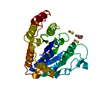
Entry
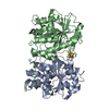
Database: PDB / ID: 2c8v
Title
Insights into the role of nucleotide-dependent conformational change in nitrogenase catalysis: Structural characterization of the nitrogenase Fe protein Leu127 deletion variant with bound MgATP
Components
NITROGENASE IRON PROTEIN 1
Keywords
OXIDOREDUCTASE / NITROGENASE / NITROGEN FIXATION / MGADP / FE PROTEIN / AV2 / 4FE- 4S / ATP-BINDING / IRON / IRON-SULFUR / METAL-BINDING / NUCLEOTIDE-BINDING
Function / homology
Function and homology information
nitrogenase / nitrogenase activity / 4 iron, 4 sulfur cluster binding / ATP binding / metal ion binding Similarity search - Function
Nitrogenase iron protein NifH / NifH/frxC family / NifH/chlL conserved site / 4Fe-4S iron sulfur cluster binding proteins, NifH/frxC family / NifH/frxC family signature 2. / NifH/frxC family signature 1. / NIFH_FRXC family profile. / P-loop containing nucleotide triphosphate hydrolases / Rossmann fold / P-loop containing nucleoside triphosphate hydrolase ...Nitrogenase iron protein NifH / NifH/frxC family / NifH/chlL conserved site / 4Fe-4S iron sulfur cluster binding proteins, NifH/frxC family / NifH/frxC family signature 2. / NifH/frxC family signature 1. / NIFH_FRXC family profile. / P-loop containing nucleotide triphosphate hydrolases / Rossmann fold / P-loop containing nucleoside triphosphate hydrolase / 3-Layer(aba) Sandwich / Alpha Beta Similarity search - Domain/homology
In the structure databanks used in Yorodumi, some data are registered as the other names, "COVID-19 virus" and "2019-nCoV". Here are the details of the virus and the list of structure data.
Jan 31, 2019. EMDB accession codes are about to change! (news from PDBe EMDB page)
EMDB accession codes are about to change! (news from PDBe EMDB page)
The allocation of 4 digits for EMDB accession codes will soon come to an end. Whilst these codes will remain in use, new EMDB accession codes will include an additional digit and will expand incrementally as the available range of codes is exhausted. The current 4-digit format prefixed with “EMD-” (i.e. EMD-XXXX) will advance to a 5-digit format (i.e. EMD-XXXXX), and so on. It is currently estimated that the 4-digit codes will be depleted around Spring 2019, at which point the 5-digit format will come into force.
The EM Navigator/Yorodumi systems omit the EMD- prefix.
Related info.:Q: What is EMD? / ID/Accession-code notation in Yorodumi/EM Navigator
Yorodumi is a browser for structure data from EMDB, PDB, SASBDB, etc.
This page is also the successor to EM Navigator detail page, and also detail information page/front-end page for Omokage search.
The word "yorodu" (or yorozu) is an old Japanese word meaning "ten thousand". "mi" (miru) is to see.
Related info.:EMDB / PDB / SASBDB / Comparison of 3 databanks / Yorodumi Search / Aug 31, 2016. New EM Navigator & Yorodumi / Yorodumi Papers / Jmol/JSmol / Function and homology information / Changes in new EM Navigator and Yorodumi
 Movie
Movie Controller

 Yorodumi
 Open data
 Basic information
 Components
 Keywords
 Function and homology information
 AZOTOBACTER VINELANDII (bacteria)
 X-RAY DIFFRACTION /
  Authors
 Citation
 Structure visualization
 Downloads & links
 Other downloads


 PDBj
PDBj


 Assembly

 Mass: 175.820 Da / Num. of mol.: 2 / Source method: obtained synthetically / Formula: Fe2S2
 Mass: 24.305 Da / Num. of mol.: 1 / Source method: obtained synthetically / Formula: Mg
 Mass: 507.181 Da / Num. of mol.: 1 / Source method: obtained synthetically / Formula: C10H16N5O13P3 / Comment: ATP, energy-carrying molecule*YM
 Mass: 18.015 Da / Num. of mol.: 99 / Source method: isolated from a natural source / Formula: H2O
 Sample preparation
 / Beamline: BL9-2 / Wavelength: 1.2
  Processing