 Movie
Movie Controller

[English] 日本語
 Yorodumi
- PDB-1fp6: THE NITROGENASE FE PROTEIN FROM AZOTOBACTER VINELANDII COMPLEXED ... -
+ Open data

- Basic information
| Entry | Database: PDB / ID: 1fp6 | ||||||
|---|---|---|---|---|---|---|---|
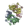
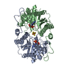
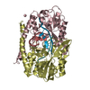
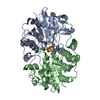
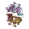
| Title | THE NITROGENASE FE PROTEIN FROM AZOTOBACTER VINELANDII COMPLEXED WITH MGADP | ||||||
 Components Components | NITROGENASE IRON PROTEIN | ||||||
 Keywords Keywords | OXIDOREDUCTASE / Nitrogenase / nitrogen fixation / nuclotide / MgADP / Fe protein / Av2 | ||||||
| Function / homology |  Function and homology information Function and homology informationnitrogenase / nitrogenase activity / 4 iron, 4 sulfur cluster binding / ATP binding / metal ion binding Similarity search - Function | ||||||
| Biological species |  Azotobacter vinelandii (bacteria) Azotobacter vinelandii (bacteria) | ||||||
| Method |  X-RAY DIFFRACTION / SYNCHROTRON / Resolution: 2.15 Å X-RAY DIFFRACTION / SYNCHROTRON / Resolution: 2.15 Å | ||||||
 Authors Authors | Jang, S.B. / Seefeldt, L.C. / Peters, J.W. | ||||||
 Citation Citation | Journal: Biochemistry / Year: 2000 Title: Insights into nucleotide signal transduction in nitrogenase: structure of an iron protein with MgADP bound. Authors: Jang, S.B. / Seefeldt, L.C. / Peters, J.W. | ||||||
| History |
|
- Structure visualization
| Structure viewer | Molecule: MolmilJmol/JSmol |
|---|
- Downloads & links
-Download
| PDBx/mmCIF format | 1fp6.cif.gz | 256.8 KB | Display | PDBx/mmCIF format |
|---|---|---|---|---|
| PDB format | pdb1fp6.ent.gz | 204.9 KB | Display | PDB format |
| PDBx/mmJSON format | 1fp6.json.gz | Tree view | PDBx/mmJSON format | |
| Others |  Other downloads Other downloads |
-Validation report
| Arichive directory | https://data.pdbj.org/pub/pdb/validation_reports/fp/1fp6ftp://data.pdbj.org/pub/pdb/validation_reports/fp/1fp6 | HTTPS FTP |
|---|
-Related structure data
| Similar structure data |
|---|

-Links
 PDBj
PDBj
- Assembly
| Deposited unit | 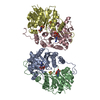
| ||||||||||
|---|---|---|---|---|---|---|---|---|---|---|---|
| 1 | 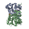
| ||||||||||
| 2 | 
| ||||||||||
| Unit cell |
| ||||||||||
| Details | homodimer complexed with two molecules of MgADP |
-Components
| #1: Protein | Mass: 31417.045 Da / Num. of mol.: 4 Source method: isolated from a genetically manipulated source Source: (gene. exp.) Azotobacter vinelandii (bacteria) / Production host: #2: Chemical | ChemComp-MG /   Mass: 24.305 Da / Num. of mol.: 4 / Source method: obtained synthetically / Formula: Mg Mass: 24.305 Da / Num. of mol.: 4 / Source method: obtained synthetically / Formula: Mg#3: Chemical |   Mass: 351.640 Da / Num. of mol.: 2 / Source method: obtained synthetically / Formula: Fe4S4 Mass: 351.640 Da / Num. of mol.: 2 / Source method: obtained synthetically / Formula: Fe4S4#4: Chemical | ChemComp-ADP /   Mass: 427.201 Da / Num. of mol.: 4 / Source method: obtained synthetically / Formula: C10H15N5O10P2 / Comment: ADP, energy-carrying molecule*YM Mass: 427.201 Da / Num. of mol.: 4 / Source method: obtained synthetically / Formula: C10H15N5O10P2 / Comment: ADP, energy-carrying molecule*YM#5: Water | ChemComp-HOH / |  Mass: 18.015 Da / Num. of mol.: 1190 / Source method: isolated from a natural source / Formula: H2O Mass: 18.015 Da / Num. of mol.: 1190 / Source method: isolated from a natural source / Formula: H2O |
|---|
-Experimental details
-Experiment
| Experiment | Method: X-RAY DIFFRACTION / Number of used crystals: 1 |
|---|
- Sample preparation
| Crystal | Density Matthews: 2.68 Å3/Da / Density % sol: 54.12 % | ||||||||||||||||||||||||||||||||||||
|---|---|---|---|---|---|---|---|---|---|---|---|---|---|---|---|---|---|---|---|---|---|---|---|---|---|---|---|---|---|---|---|---|---|---|---|---|---|
| Crystal grow | Temperature: 298 K / Method: liquid diffusion / pH: 8.5 Details: PEG 4000, 0.1M Tris HCl, Sodium Acetate, Glycerol, pH 8.5, LIQUID DIFFUSION, temperature 298K | ||||||||||||||||||||||||||||||||||||
| Crystal grow | *PLUS Method: batch method | ||||||||||||||||||||||||||||||||||||
| Components of the solutions | *PLUS
|
-Data collection
| Diffraction | Mean temperature: 100 K |
|---|---|
| Diffraction source | Source: SYNCHROTRON / Site: SSRL  / Beamline: BL9-1 / Wavelength: 0.97 / Beamline: BL9-1 / Wavelength: 0.97 |
| Detector | Type: MARRESEARCH / Detector: IMAGE PLATE / Date: Feb 24, 2000 |
| Radiation | Protocol: SINGLE WAVELENGTH / Monochromatic (M) / Laue (L): M / Scattering type: x-ray |
| Radiation wavelength | Wavelength: 0.97 Å / Relative weight: 1 |
| Reflection | Resolution: 2.15→30 Å / Num. all: 65315 / Num. obs: 65315 / % possible obs: 88.8 % / Redundancy: 3.2 % / Rmerge(I) obs: 0.064 / Net I/σ(I): 7.7 |
| Reflection shell | Resolution: 2.15→2.2 Å / Redundancy: 2.9 % / Rmerge(I) obs: 0.279 / Num. unique all: 3603 / % possible all: 69.4 |
| Reflection | *PLUS Num. measured all: 210392 |
| Reflection shell | *PLUS % possible obs: 69.4 % / Mean I/σ(I) obs: 2.6 |
- Processing
| Software |
| |||||||||||||||||||||||||
|---|---|---|---|---|---|---|---|---|---|---|---|---|---|---|---|---|---|---|---|---|---|---|---|---|---|---|
| Refinement | Resolution: 2.15→30 Å / Cross valid method: THROUGHOUT / σ(F): 1 / Stereochemistry target values: Engh & Huber
| |||||||||||||||||||||||||
| Refinement step | Cycle: LAST / Resolution: 2.15→30 Å
| |||||||||||||||||||||||||
| Refine LS restraints |
| |||||||||||||||||||||||||
| Software | *PLUS Name: X-PLOR / Version: 3.851 / Classification: refinement | |||||||||||||||||||||||||
| Refinement | *PLUS σ(F): 1 / % reflection Rfree: 5 % / Rfactor obs: 0.2 / Rfactor Rwork: 0.2 | |||||||||||||||||||||||||
| Solvent computation | *PLUS | |||||||||||||||||||||||||
| Displacement parameters | *PLUS |
