 ムービー
ムービー コントローラー


+ データを開く

- 基本情報
基本情報
| 登録情報 | データベース: PDB / ID: 1de0 | ||||||
|---|---|---|---|---|---|---|---|
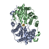
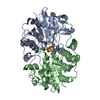
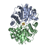
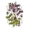
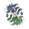
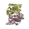
| タイトル | MODULATING THE MIDPOINT POTENTIAL OF THE [4FE-4S] CLUSTER OF THE NITROGENASE FE PROTEIN | ||||||
 要素 要素 | NITROGENASE IRON PROTEIN | ||||||
 キーワード キーワード | OXIDOREDUCTASE / REDOX PROTEINS / [FES] CLUSTERS / FE PROTEIN | ||||||
| 機能・相同性 |  機能・相同性情報 機能・相同性情報nitrogenase / nitrogenase activity / 4 iron, 4 sulfur cluster binding / ATP binding / metal ion binding 類似検索 - 分子機能 | ||||||
| 生物種 |  Azotobacter vinelandii (窒素固定) Azotobacter vinelandii (窒素固定) | ||||||
| 手法 |  X線回折 / 解像度: 2.4 Å X線回折 / 解像度: 2.4 Å | ||||||
 データ登録者 データ登録者 | Jang, S.B. / Seefeldt, L.C. / Peters, J.W. | ||||||
 引用 引用 | ジャーナル: Biochemistry / 年: 2000 タイトル: Modulating the midpoint potential of the [4Fe-4S] cluster of the nitrogenase Fe protein. 著者: Jang, S.B. / Seefeldt, L.C. / Peters, J.W. #1: ジャーナル: Science / 年: 1992タイトル: Crystallographic Structure of the Nitrogenase Iron Protein from Azotobacter vinelandii 著者: Georgiadis, M.M. / Komiya, H. / Chakrabarti, P. / Woo, D. / Kornuc, J.J. / Rees, D.C. #2: ジャーナル: J.Mol.Biol. / 年: 1998タイトル: Conformational Variability in Structures of the Nitrogenase Iron Proteins from Azotobacter vinelandii and Clostridium pasteurianum 著者: Schlessman, J.L. / Woo, D. / Joshua-Tor, L. / Howard, J.B. / Rees, D.C. #3: ジャーナル: Nature / 年: 1997タイトル: Structure of ADP x AlF4(-)-stabilized Nitrogenase Complex and its Implications for Signal Transduction 著者: Schindelin, H. / Kisker, C. / Schlessman, J.L. / Howard, J.B. / Rees, D.C. | ||||||
| 履歴 |
|
- 構造の表示
構造の表示
| 構造ビューア | 分子: MolmilJmol/JSmol |
|---|
- ダウンロードとリンク
-ダウンロード
| PDBx/mmCIF形式 | 1de0.cif.gz | 124.1 KB | 表示 | PDBx/mmCIF形式 |
|---|---|---|---|---|
| PDB形式 | pdb1de0.ent.gz | 96.6 KB | 表示 | PDB形式 |
| PDBx/mmJSON形式 | 1de0.json.gz | ツリー表示 | PDBx/mmJSON形式 | |
| その他 |  その他のダウンロード その他のダウンロード |
-検証レポート
| アーカイブディレクトリ | https://data.pdbj.org/pub/pdb/validation_reports/de/1de0ftp://data.pdbj.org/pub/pdb/validation_reports/de/1de0 | HTTPS FTP |
|---|
-関連構造データ
| 関連構造データ | |
|---|---|
| 類似構造データ |


-リンク
 PDBj
PDBj
- 集合体
集合体
| 登録構造単位 | 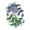
| ||||||||
|---|---|---|---|---|---|---|---|---|---|
| 1 |
| ||||||||
| 単位格子 |
|
-要素
| #1: タンパク質 | 分子量: 31456.084 Da / 分子数: 2 / 変異: F135W / 由来タイプ: 天然 / 由来: (天然) Azotobacter vinelandii (窒素固定) / 参照: UniProt: P00459, nitrogenase#2: 化合物 | ChemComp-SF4 / |   分子量: 351.640 Da / 分子数: 1 / 由来タイプ: 合成 / 式: Fe4S4 分子量: 351.640 Da / 分子数: 1 / 由来タイプ: 合成 / 式: Fe4S4#3: 水 | ChemComp-HOH / |  分子量: 18.015 Da / 分子数: 224 / 由来タイプ: 天然 / 式: H2O 分子量: 18.015 Da / 分子数: 224 / 由来タイプ: 天然 / 式: H2O |
|---|
-実験情報
-実験
| 実験 | 手法: X線回折 / 使用した結晶の数: 1 |
|---|
- 試料調製
試料調製
| 結晶 | マシュー密度: 2.55 Å3/Da / 溶媒含有率: 51.83 % | ||||||||||||||||||||
|---|---|---|---|---|---|---|---|---|---|---|---|---|---|---|---|---|---|---|---|---|---|
| 結晶化 | 温度: 277 K / 手法: small tubes / pH: 8.5 詳細: PEG 4000, 0.1 M TRIS HCL, 0.2 M SODIUM ACETATE, pH 8.5, SMALL TUBES, temperature 277K | ||||||||||||||||||||
| 結晶化 | *PLUS 手法: batch method | ||||||||||||||||||||
| 溶液の組成 | *PLUS
|
-データ収集
| 回折 | 平均測定温度: 100 K |
|---|---|
| 放射光源 | 由来: 回転陽極 / タイプ: RIGAKU RU300 / 波長: 1.5418 |
| 検出器 | タイプ: RIGAKU RAXIS IIC / 検出器: IMAGE PLATE / 日付: 1999年2月15日 |
| 放射 | プロトコル: SINGLE WAVELENGTH / 単色(M)・ラウエ(L): M / 散乱光タイプ: x-ray |
| 放射波長 | 波長: 1.5418 Å / 相対比: 1 |
| 反射 | 解像度: 2.4→20 Å / Num. all: 265157 / Num. obs: 24852 / % possible obs: 9.37 % / Observed criterion σ(F): 0 / Observed criterion σ(I): 18.4 / 冗長度: 6 % / Biso Wilson estimate: 36.3 Å2 / Rmerge(I) obs: 0.077 |
| 反射 シェル | 解像度: 2.4→2.44 Å / 冗長度: 6 % / Rmerge(I) obs: 0.253 / % possible all: 0.2 |
| 反射 | *PLUS % possible obs: 95.2 % / Num. measured all: 265157 |
| 反射 シェル | *PLUS % possible obs: 75.5 % / Mean I/σ(I) obs: 3.9 |
- 解析
解析
| ソフトウェア |
| ||||||||||||||||||||||||||||||||||||||||||||||||||||||||||||
|---|---|---|---|---|---|---|---|---|---|---|---|---|---|---|---|---|---|---|---|---|---|---|---|---|---|---|---|---|---|---|---|---|---|---|---|---|---|---|---|---|---|---|---|---|---|---|---|---|---|---|---|---|---|---|---|---|---|---|---|---|---|
| 精密化 | 解像度: 2.4→20 Å / σ(F): 0 / σ(I): 0
| ||||||||||||||||||||||||||||||||||||||||||||||||||||||||||||
| 精密化ステップ | サイクル: LAST / 解像度: 2.4→20 Å
| ||||||||||||||||||||||||||||||||||||||||||||||||||||||||||||
| 拘束条件 |
| ||||||||||||||||||||||||||||||||||||||||||||||||||||||||||||
| ソフトウェア | *PLUS 名称: X-PLOR / バージョン: 3.1 / 分類: refinement | ||||||||||||||||||||||||||||||||||||||||||||||||||||||||||||
| 精密化 | *PLUS | ||||||||||||||||||||||||||||||||||||||||||||||||||||||||||||
| 溶媒の処理 | *PLUS | ||||||||||||||||||||||||||||||||||||||||||||||||||||||||||||
| 原子変位パラメータ | *PLUS Biso mean: 50.3 Å2 | ||||||||||||||||||||||||||||||||||||||||||||||||||||||||||||
| 拘束条件 | *PLUS タイプ: x_angle_deg / Dev ideal: 2.576 |
