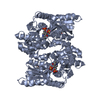
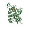
Deposited unit
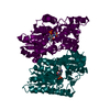
A: 1-aminocyclopropane-1-carboxylate deaminase
B: 1-aminocyclopropane-1-carboxylate deaminase
C: 1-aminocyclopropane-1-carboxylate deaminase
D: 1-aminocyclopropane-1-carboxylate deaminase
E: 1-aminocyclopropane-1-carboxylate deaminase
F: 1-aminocyclopropane-1-carboxylate deaminase
G: 1-aminocyclopropane-1-carboxylate deaminase
H: 1-aminocyclopropane-1-carboxylate deaminase
I: 1-aminocyclopropane-1-carboxylate deaminase
J: 1-aminocyclopropane-1-carboxylate deaminase
K: 1-aminocyclopropane-1-carboxylate deaminase
L: 1-aminocyclopropane-1-carboxylate deaminase
M: 1-aminocyclopropane-1-carboxylate deaminase
N: 1-aminocyclopropane-1-carboxylate deaminase
O: 1-aminocyclopropane-1-carboxylate deaminase
P: 1-aminocyclopropane-1-carboxylate deaminase
Q: 1-aminocyclopropane-1-carboxylate deaminase
R: 1-aminocyclopropane-1-carboxylate deaminase
S: 1-aminocyclopropane-1-carboxylate deaminase
T: 1-aminocyclopropane-1-carboxylate deaminase
U: 1-aminocyclopropane-1-carboxylate deaminase
V: 1-aminocyclopropane-1-carboxylate deaminase
W: 1-aminocyclopropane-1-carboxylate deaminase
X: 1-aminocyclopropane-1-carboxylate deaminase
hetero molecules Summary Component details
Theoretical mass Number of molelcules Total (without water) 853,562 48 Polymers 845,588 24 Non-polymers 7,974 24 Water 14,484 804
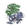
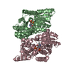
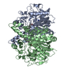
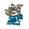
1
A: 1-aminocyclopropane-1-carboxylate deaminase
B: 1-aminocyclopropane-1-carboxylate deaminase
hetero molecules Summary Component details Symmetry operations Calculated values
Theoretical mass Number of molelcules Total (without water) 71,130 4 Polymers 70,466 2 Non-polymers 664 2 Water 36 2
Type Name Symmetry operation Number identity operation 1_555 x,y,z 1
Buried area 4670 Å2 ΔGint -27 kcal/mol Surface area 22930 Å2 Method
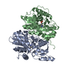
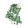
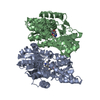
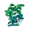
2
C: 1-aminocyclopropane-1-carboxylate deaminase
D: 1-aminocyclopropane-1-carboxylate deaminase
hetero molecules Summary Component details Symmetry operations Calculated values
Theoretical mass Number of molelcules Total (without water) 71,130 4 Polymers 70,466 2 Non-polymers 664 2 Water 36 2
Type Name Symmetry operation Number identity operation 1_555 x,y,z 1
Buried area 4720 Å2 ΔGint -27 kcal/mol Surface area 22900 Å2 Method
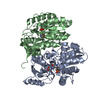
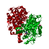
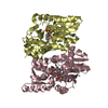
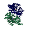
3
E: 1-aminocyclopropane-1-carboxylate deaminase
F: 1-aminocyclopropane-1-carboxylate deaminase
hetero molecules Summary Component details Symmetry operations Calculated values
Theoretical mass Number of molelcules Total (without water) 71,130 4 Polymers 70,466 2 Non-polymers 664 2 Water 36 2
Type Name Symmetry operation Number identity operation 1_555 x,y,z 1
Buried area 4680 Å2 ΔGint -28 kcal/mol Surface area 23170 Å2 Method
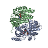
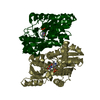
4
G: 1-aminocyclopropane-1-carboxylate deaminase
H: 1-aminocyclopropane-1-carboxylate deaminase
hetero molecules Summary Component details Symmetry operations Calculated values
Theoretical mass Number of molelcules Total (without water) 71,130 4 Polymers 70,466 2 Non-polymers 664 2 Water 36 2
Type Name Symmetry operation Number identity operation 1_555 x,y,z 1
Buried area 4680 Å2 ΔGint -25 kcal/mol Surface area 23030 Å2 Method
5
I: 1-aminocyclopropane-1-carboxylate deaminase
J: 1-aminocyclopropane-1-carboxylate deaminase
hetero molecules Summary Component details Symmetry operations Calculated values
Theoretical mass Number of molelcules Total (without water) 71,130 4 Polymers 70,466 2 Non-polymers 664 2 Water 36 2
Type Name Symmetry operation Number identity operation 1_555 x,y,z 1
Buried area 4770 Å2 ΔGint -26 kcal/mol Surface area 23010 Å2 Method
6
K: 1-aminocyclopropane-1-carboxylate deaminase
L: 1-aminocyclopropane-1-carboxylate deaminase
hetero molecules Summary Component details Symmetry operations Calculated values
Theoretical mass Number of molelcules Total (without water) 71,130 4 Polymers 70,466 2 Non-polymers 664 2 Water 36 2
Type Name Symmetry operation Number identity operation 1_555 x,y,z 1
Buried area 4690 Å2 ΔGint -27 kcal/mol Surface area 22880 Å2 Method
7
M: 1-aminocyclopropane-1-carboxylate deaminase
N: 1-aminocyclopropane-1-carboxylate deaminase
hetero molecules Summary Component details Symmetry operations Calculated values
Theoretical mass Number of molelcules Total (without water) 71,130 4 Polymers 70,466 2 Non-polymers 664 2 Water 36 2
Type Name Symmetry operation Number identity operation 1_555 x,y,z 1
Buried area 4710 Å2 ΔGint -28 kcal/mol Surface area 23000 Å2 Method
8
O: 1-aminocyclopropane-1-carboxylate deaminase
P: 1-aminocyclopropane-1-carboxylate deaminase
hetero molecules Summary Component details Symmetry operations Calculated values
Theoretical mass Number of molelcules Total (without water) 71,130 4 Polymers 70,466 2 Non-polymers 664 2 Water 36 2
Type Name Symmetry operation Number identity operation 1_555 x,y,z 1
Buried area 4770 Å2 ΔGint -27 kcal/mol Surface area 23010 Å2 Method
9
Q: 1-aminocyclopropane-1-carboxylate deaminase
R: 1-aminocyclopropane-1-carboxylate deaminase
hetero molecules Summary Component details Symmetry operations Calculated values
Theoretical mass Number of molelcules Total (without water) 71,130 4 Polymers 70,466 2 Non-polymers 664 2 Water 36 2
Type Name Symmetry operation Number identity operation 1_555 x,y,z 1
Buried area 4710 Å2 ΔGint -27 kcal/mol Surface area 23050 Å2 Method
10
S: 1-aminocyclopropane-1-carboxylate deaminase
T: 1-aminocyclopropane-1-carboxylate deaminase
hetero molecules Summary Component details Symmetry operations Calculated values
Theoretical mass Number of molelcules Total (without water) 71,130 4 Polymers 70,466 2 Non-polymers 664 2 Water 36 2
Type Name Symmetry operation Number identity operation 1_555 x,y,z 1
Buried area 4750 Å2 ΔGint -28 kcal/mol Surface area 23160 Å2 Method
11
U: 1-aminocyclopropane-1-carboxylate deaminase
V: 1-aminocyclopropane-1-carboxylate deaminase
hetero molecules Summary Component details Symmetry operations Calculated values
Theoretical mass Number of molelcules Total (without water) 71,130 4 Polymers 70,466 2 Non-polymers 664 2 Water 36 2
Type Name Symmetry operation Number identity operation 1_555 x,y,z 1
Buried area 4680 Å2 ΔGint -28 kcal/mol Surface area 22950 Å2 Method
12
W: 1-aminocyclopropane-1-carboxylate deaminase
X: 1-aminocyclopropane-1-carboxylate deaminase
hetero molecules Summary Component details Symmetry operations Calculated values
Theoretical mass Number of molelcules Total (without water) 71,130 4 Polymers 70,466 2 Non-polymers 664 2 Water 36 2
Type Name Symmetry operation Number identity operation 1_555 x,y,z 1
Buried area 4730 Å2 ΔGint -27 kcal/mol Surface area 23030 Å2 Method
Unit cell Length a, b, c (Å) 105.87, 147.28, 149.07 Angle α, β, γ (deg.) 73.18, 90.11, 68.49 Int Tables number 1 Space group name H-M P1
 Movie
Movie Controller

 Yorodumi
 Open data
 Basic information
 Components
 Keywords
 Function and homology information
 Pyrococcus horikoshii (archaea)
 X-RAY DIFFRACTION /
  Authors
 Citation
 Structure visualization
 Downloads & links
 Other downloads


 PDBj
PDBj
 Assembly


 Mass: 332.246 Da / Num. of mol.: 24 / Source method: obtained synthetically / Formula: C12H17N2O7P
 Mass: 18.015 Da / Num. of mol.: 804 / Source method: isolated from a natural source / Formula: H2O
 Sample preparation
 / Beamline: BL41XU / Wavelength: 1 Å
 Processing